Diabetes-induced fetal growth retardation is associated with suppression of NF-kappaB activity in embryos
- PMID: 17491656
- PMCID: PMC1762494
- DOI: 10.1900/RDS.2005.2.27
Diabetes-induced fetal growth retardation is associated with suppression of NF-kappaB activity in embryos
Abstract
Background: Mechanisms underlying diabetes-induced fetal growth retardation remain largely undefined. Two events such as the persistent activation of apoptosis or suppression of cell proliferation in embryos might directly result in fetal growth retardation. Evidence implicating the transcription factor NF-kappaB in the regulation of the physiological and teratogen-induced apoptosis as well as cell proliferation suggests that it may be a component of mechanisms underlying this pathology. To address this issue, this study was designed to test: 1) whether diabetes-induced fetal growth retardation is preceded by the modulation of NF-kappaB activity in embryos at the late stage of organogenesis and 2) whether apoptosis is altered in these embryos.
Methods: The embryos and placentas of streptozotocin-induced diabetic mice collected on days 13 and 15 of pregnancy were used to evaluate the expression of NF-kappaB, IkappaBalpha and phosphorylated (p)-IkappaBalpha proteins by Western blot analysis and NF-kappaB DNA binding by an ELISA-based method. The detection of apoptotic cells was performed by the TUNEL assay and the expression of a proapoptotic protein Bax was evaluated by the Western blot.
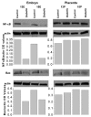
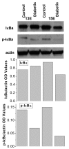
Results: The embryos of diabetic mice were significantly growth retarded, whereas the placental weight did not differ in diabetic or control females. Levels of NF-kappaB and p-IkappaBalpha proteins as well as the amount of NF-kappaB DNA binding was lower in embryos of diabetic mice as compared to those in controls. However, neither excessive apoptosis nor an increased Bax expression was found in growth-retarded embryos and their placentas.
Conclusion: The study herein revealed that diabetes-induced fetal growth retardation is associated with the suppression of NF-kappaB activity in embryos, which seems to be realized at the level of IkappaB degradation.
Figures


References
-
- Eriksson UJ, Borg LA, Forsberg H, Styrud J. Diabetic embryopathy. Studies with animal and in vitro models. Diabetes. 1991;40(Suppl 2):94–98. - PubMed
-
- Reece EA, Homko CJ. Multifactorial basis of the syndrome of diabetic embryopathy. Teratology. 1996;54:171–182. - PubMed
-
- Moley KH. Hyperglycemia and apoptosis: mechanisms for congenital malformation and pregnancy loss in diabetic women. Trends Endocrinol Metab. 2001;12:78–82. - PubMed
-
- Sun F, Kawasaki E, Akazawa S, Hishikawa Y, Sugahara K, Kamihira S, Koji T, Eguchi K. Apoptosis and its pathway in early post-implantation embryos of diabetic rats. Diabetes Res Clin Pract. 2005;67:110–118. - PubMed
-
- Chen FE, Ghosh G. Regulation of DNA binding by Rel/NF-kappaB transcription factors: structural views. Oncogene. 1999;18:6845–6852. - PubMed
LinkOut - more resources
Full Text Sources
Other Literature Sources
Molecular Biology Databases
Research Materials
