. 2007 Jun;117(6):1699-707.
doi: 10.1172/JCI30575. Epub 2007 May 10.

Mice lacking the signaling molecule CalDAG-GEFI represent a model for leukocyte adhesion deficiency type III

Affiliations

Mice lacking the signaling molecule CalDAG-GEFI represent a model for leukocyte adhesion deficiency type III

Wolfgang Bergmeier et al. J Clin Invest. 2007 Jun.

Abstract

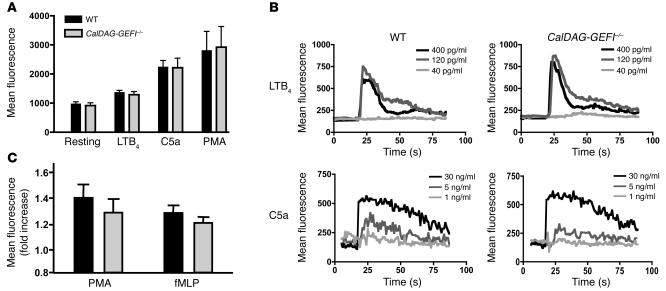
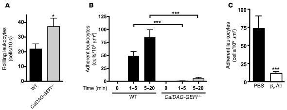
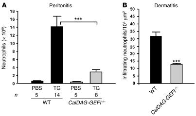
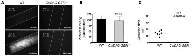
Single gene mutations in beta integrins can account for functional defects of individual cells of the hematopoietic system. In humans, mutations in beta(2) integrin lead to leukocyte adhesion deficiency (LAD) syndrome and mutations in beta(3) integrin cause the bleeding disorder Glanzmann thrombasthenia. However, multiple defects in blood cells involving various beta integrins (beta(1), beta(2), and beta(3)) occur simultaneously in patients with the recently described LAD type III (LAD-III). Here we show that the product of a single gene, Ca(2+) and diacylglycerol-regulated guanine nucleotide exchange factor I (CalDAG-GEFI), controlled the activation of all 3 integrins in the hematopoietic system. Neutrophils from CalDAG-GEFI(-/-) mice exhibited strong defects in Rap1 and beta(1) and beta(2) integrin activation while maintaining normal calcium flux, degranulation, and ROS generation. Neutrophils from CalDAG-GEFI-deficient mice failed to adhere firmly to stimulated venules and to migrate into sites of inflammation. Furthermore, CalDAG-GEFI regulated the activation of beta(1) and beta(3) integrins in platelets, and CalDAG-GEFI deficiency caused complete inhibition of arterial thrombus formation in mice. Thus, mice engineered to lack CalDAG-GEFI have a combination of defects in leukocyte and platelet functions similar to that of LAD-III patients.


Figures

Figure 1
Figure 1. Degranulation, calcium flux, and ROS production in stimulated neutrophils.
(A) Mac-1 expression. PBLs from WT and CalDAG-GEFI–/– mice were kept resting or were activated for 10 minutes with LTB4 (300 nM), C5a (50 ng/ml), or PMA (200 nM), stained with antibodies against Mac-1, and immediately analyzed by flow cytometry. n = 6. (B) Calcium flux. PBLs from WT and CalDAG-GEFI–/– mice were loaded with the calcium-sensitive dye Fluo-3, activated with the indicated doses of LTB4 or C5a, and immediately analyzed by flow cytometry. Results are representative of 6 individual experiments. (C) ROS formation. PBLs from WT and CalDAG-GEFI–/– mice were loaded with the ROS-sensitive agent DCFDA, activated with PMA (2 μM) or fMLP (5 μM) for 30 minutes at 37°C, and immediately analyzed by flow cytometry. n = 5. ROS production is expressed as fold increase of mean fluorescence intensity over unstimulated cells. No significant differences in Mac-1 expression, calcium flux, or ROS production were observed between WT and CalDAG-GEFI–deficient neutrophils.
Figure 2
Figure 2. Impaired Rap1 activation in CalDAG-GEFI–deficient neutrophils.
Western blots of affinity-precipitated Rap1-GTP showing strongly decreased Rap1 activation in CalDAG-GEFI–deficient neutrophils (–/–) stimulated with LTB4 (300 nM for 30 seconds), C5a (75 ng/ml for 30 seconds), or PAF (3 or 30 nM for 30 seconds) relative to that of WT neutrophils (+/+). Equivalent loading of GTP onto Rap1 in neutrophils from WT and CalDAG-GEFI–/– mice was shown by preincubation of lysates with GTP-γS. Total Rap1 levels were determined in whole cell lysates of WT and CalDAG-GEFI–deficient neutrophils. Results are representative of 3 individual experiments.
Figure 3
Figure 3. CalDAG-GEFI deficiency causes impaired β1 integrin– and β2 integrin–mediated adhesion of neutrophils in vitro.
(A and B) Neutrophil adhesion to fibronectin (A) or fibrinogen (B) in vitro. WT or CalDAG-GEFI–deficient neutrophils isolated from bone marrow were added to fibronectin- or fibrinogen-coated plates and incubated for 30 minutes in the presence or absence (i.e., resting) of 300 nM LTB4 or 3 or 30 nM PAF. Plates were washed and adherent neutrophils were counted. n = 4. *P < 0.05, **P < 0.01, ***P < 0.001.
Figure 4
Figure 4. Impaired firm adhesion of leukocytes to mesenteric venules in CalDAG-GEFI–/– mice.
(A) Leukocyte rolling in vivo. The number of rolling leukocytes in WT or CalDAG-GEFI–/– mice was determined by intravital microscopy. Mice were infused with rhodamine 6G to label circulating leukocytes. Leukocyte rolling was quantified 1–5 minutes after superfusion with 300 nM LTB4. (B) Firm adhesion. WT or CalDAG-GEFI–deficient leukocytes were considered firmly adherent when they remained stationary for more than 30 seconds. n = 6. (C) Blocking antibodies to β2 integrin markedly reduce leukocyte adhesion to mesenteric venules. Leukocyte adhesion within the first 5 minutes after LTB4 superfusion was studied in WT mice infused with PBS or 40 μg anti-β2 antibodies (β2 Ab). n = 3. *P < 0.05, ***P < 0.001. No significant difference in leukocyte adhesion was observed between untreated and control IgG–treated WT mice (not shown).
Figure 5
Figure 5. CalDAG-GEFI regulates neutrophil extravasation.
(A) TG-induced peritonitis. Infiltrating leukocytes were isolated from the peritoneum of WT and CalDAG-GEFI–/– mice 4 hours after injection of PBS or 3% TG. Neutrophil counts were determined by morphological analyses of Diff-Quik–stained cytocentrifuge preparations by an observer blinded to the genotype. (B) Croton oil–induced dermatitis. Ears of WT and CalDAG-GEFI–/– mice were painted with croton oil, and infiltrating neutrophils were counted 6 hours later in H&E-stained ear sections. Few neutrophils were found in vehicle-treated ears. n = 3–5. ***P < 0.001.
Figure 6
Figure 6. Impaired activation of β1 integrins in CalDAG-GEFI–deficient platelets.
(A and B) Biotinylated WT and CalDAG-GEFI–deficient platelets were stimulated with PAR4p (2 mM) or U46619/ADP (5 or 10 μM) in the presence of a blocking antibody to αIIbβ3 and allowed to adhere for 30 minutes under static conditions to laminin (A) or fibronectin (B) in microtiter plates. A separate group of WT platelets was pretreated with a blocking antibody to α6 (adhesion to laminin) or with EDTA (adhesion to fibronectin) to demonstrate the integrin dependency of the adhesion process. Adherent platelets were quantified colorimetrically for peroxidase activity. Data are mean ± SEM of 3 individual experiments in triplicate wells. **P < 0.01, ***P < 0.001. Similar results were observed with nonbiotinylated platelets when the number of adhesive platelets was determined by light microscopy (not shown).
Figure 7
Figure 7. Platelet adhesion and thrombus formation in FeCl3-injured arterioles.
(A) WT and CalDAG-GEFI–/– mice were injected with calcein-green-labeled platelets of the respective genotype, and platelet adhesion was monitored in mesenteric arterioles (diameter, WT, 90.7 ± 3.5 μm; CalDAG-GEFI–/–, 96.4 ± 4.0 μm) upon application of FeCl3. Images show platelet adhesion and thrombus formation in arterioles at the indicated times after application of FeCl3. The direction of blood flow is from top to bottom. (B and C) Comparison of the number of tethering platelets (B) and the time of occlusion (C) in FeCl3-treated mesenteric arterioles of WT and CalDAG-GEFI–/– mice. No thrombi formed in the mutant mice during the 40-minute observation period. ***P < 0.001.

References

    1. Hynes R.O. Integrins: bidirectional, allosteric signaling machines. Cell. 2002;110:673–687. - PubMed
    1. Sims T.N., Dustin M.L. The immunological synapse: integrins take the stage. Immunol. Rev. 2002;186:100–117. - PubMed
    1. Simon S.I., Green C.E. Molecular mechanics and dynamics of leukocyte recruitment during inflammation. Annu. Rev. Biomed. Eng. 2005;7:151–185. - PubMed
    1. Savage B., Cattaneo M., Ruggeri Z.M. Mechanisms of platelet aggregation. Curr. Opin. Hematol. 2001;8:270–276. - PubMed
    1. Anderson D.C., Springer T.A. Leukocyte adhesion deficiency: an inherited defect in the Mac-1, LFA-1, and p150,95 glycoproteins. Annu. Rev. Med. 1987;38:175–194. - PubMed

Publication types

MeSH terms