. 2007 Jun;117(6):1538-49.
doi: 10.1172/JCI30634. Epub 2007 May 10.

IL-33 and ST2 comprise a critical biomechanically induced and cardioprotective signaling system

Affiliations

IL-33 and ST2 comprise a critical biomechanically induced and cardioprotective signaling system

Shoji Sanada et al. J Clin Invest. 2007 Jun.

Abstract

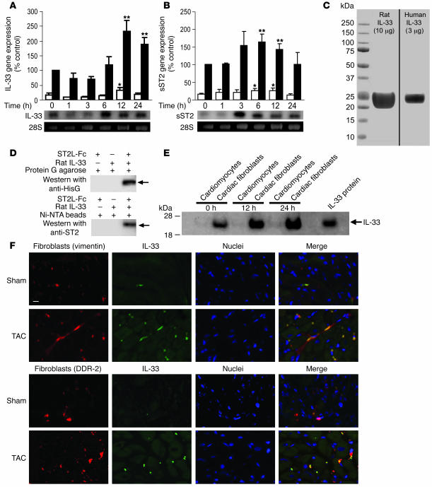
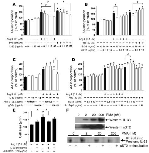
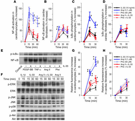
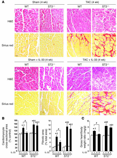
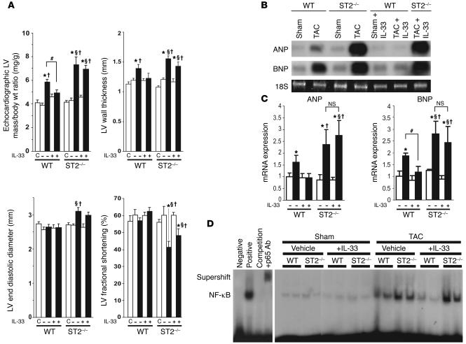
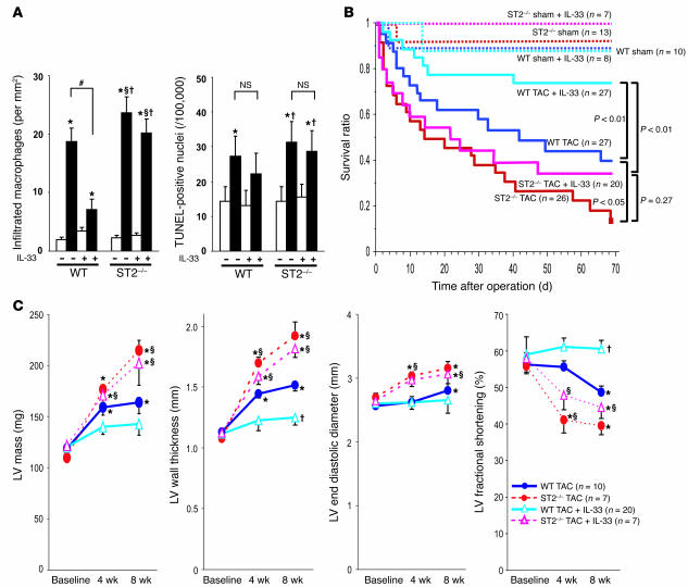
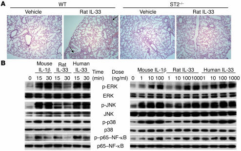
ST2 is an IL-1 receptor family member with transmembrane (ST2L) and soluble (sST2) isoforms. sST2 is a mechanically induced cardiomyocyte protein, and serum sST2 levels predict outcome in patients with acute myocardial infarction or chronic heart failure. Recently, IL-33 was identified as a functional ligand of ST2L, allowing exploration of the role of ST2 in myocardium. We found that IL-33 was a biomechanically induced protein predominantly synthesized by cardiac fibroblasts. IL-33 markedly antagonized angiotensin II- and phenylephrine-induced cardiomyocyte hypertrophy. Although IL-33 activated NF-kappaB, it inhibited angiotensin II- and phenylephrine-induced phosphorylation of inhibitor of NF-kappa B alpha (I kappa B alpha) and NF-kappaB nuclear binding activity. sST2 blocked antihypertrophic effects of IL-33, indicating that sST2 functions in myocardium as a soluble decoy receptor. Following pressure overload by transverse aortic constriction (TAC), ST2(-/-) mice had more left ventricular hypertrophy, more chamber dilation, reduced fractional shortening, more fibrosis, and impaired survival compared with WT littermates. Furthermore, recombinant IL-33 treatment reduced hypertrophy and fibrosis and improved survival after TAC in WT mice, but not in ST2(-/-) littermates. Thus, IL-33/ST2 signaling is a mechanically activated, cardioprotective fibroblast-cardiomyocyte paracrine system, which we believe to be novel. IL-33 may have therapeutic potential for beneficially regulating the myocardial response to overload.


Figures

Figure 1
Figure 1. IL-33 is induced by mechanical strain in cardiac fibroblasts.
(A and B) Quantitative analyses of gene expression of IL-33 by quantitative PCR (A) and sST2 by Northern analysis (B) in rat neonatal cardiomyocytes (white bars) and fibroblasts (black bars) are shown above with representative images from Northern analyses of cardiac fibroblast RNA. Cells were subjected to cyclic strain (8%, 1 Hz) for the indicated periods. Values are relative to β-tubulin expression and are expressed as percentage of control in cardiac fibroblasts. Data are from at least 3 sets of independent experiments. *P < 0.05, **P < 0.01 versus baseline. (C) Coomassie stain showed that the recombinant mature rat and human IL-33 with N-terminal His tag (10 and 3 μg protein, respectively, was loaded) were of high purity. (D) Pull-down assay of recombinant rat IL-33 with mouse ST2L-Fc protein. The recombinant protein exhibited specific binding to mouse ST2. (E) Western analysis of cardiomyocytes and cardiac fibroblasts subjected to cyclic strain (each 10 μg protein sample from whole cell lysate) for the indicated periods. For reference, 0.1 ng of recombinant IL-33 was applied in the right lane. (F) Representative immunofluorescence microscopy images of left ventricular samples 1 week after sham operation or TAC. Anti-vimentin (top panels) or anti–discoidin domain receptor–2 (DDR-2; bottom panels) antibody was used to detect fibroblasts (red) for dual staining with IL-33 (green). Pressure overload by TAC induced IL-33 expression, particularly in noncardiomyocyte interstitial cells. Scale bar: 10 μm.
Figure 2
Figure 2. IL-33 blocks prohypertrophic stimuli in cardiomyocytes and sST2 inhibits IL-33.
(A) IL-33 demonstrated a nonsignificant trend toward stimulating hypertrophy, but IL-33 blocked angiotensin II– and phenylephrine-induced (Phe) leucine uptake in a dose-dependent manner. (B) sST2 dose-dependently reversed the antihypertrophic effect of IL-33 under angiotensin II and phenylephrine. (C) Anti-ST2L monoclonal antibody, which blocks membrane-bound ST2L receptor–binding activity, blocked the antihypertrophic effect of IL-33, unlike control IgG. (D) Leucine uptake was not affected by sST2 compared with either baseline or control protein IL-1R–related protein 2 (IL-1Rrp2), but further enhanced hypertrophy under angiotensin II and phenylephrine. Data are from 3–5 sets of experiments. (E) Quantitative analysis of in vitro cell size measurements of cardiomyocytes was consistent with leucine incorporation assays (n = 200 each). *P < 0.05 versus baseline; #P < 0.05. (F) Induced secretion of both sST2 and IL-33 can reduce free IL-33. Cardiac fibroblasts were treated with indicated doses of PMA for 24 hours to induce sST2 and IL-33. In the top blots, conditioned media (20 μl) were analyzed by Western analysis. PMA dose-dependently increased secretion of both IL-33 and sST2. Below, media were preincubated in the presence or absence of 20 μg sST2-Fc protein and then incubated with presaturated beads for 2 hours. Samples preincubated with sST2-Fc had little IL-33, indicating that preincubation with sST2-Fc removed free IL-33. PMA dose-dependently decreased free IL-33 despite an increase in overall IL-33; these data suggest that induced sST2 can function as a decoy receptor, decreasing free IL-33.
Figure 3
Figure 3. IL-33 transiently activates NF-κB but blocks NF-κB activation by hypertrophic stimuli.
(A and B) NF-κB nuclear binding activity measured by EMSA in cardiomyocytes (A) and cardiac fibroblasts (B). (C and D) IκBα phosphorylation evaluated by Western analysis in cardiomyocytes (C) and cardiac fibroblasts (D). Values are relative to control density and are expressed as percent increase compared with control. Both angiotensin II and phenylephrine significantly activated NF-κB. IL-33 also activated NF-κB, but IL-33 markedly attenuated angiotensin II– and phenylephrine-induced NF-κB activation in cardiomyocytes, unlike in cardiac fibroblasts. IκBα phosphorylation was similarly affected by IL-33 treatment. (E) IL-33 (10 ng/ml) did not block IκBα phosphorylation (Western analysis) and NF-κB activity (EMSA) induced by PDGF-BB (10 ng/ml) or TNF-α (10 ng/ml), unlike angiotensin II and phenylephrine. (F) Western analysis for MAPKs and Akt in cardiomyocytes. IL-33 (10 ng/ml) activated all MAPKs, generally to a lesser extent than did IL-1β (10 ng/ml). IL-33 attenuated angiotensin II–induced phosphorylation of p38 MAPK and JNK, but not ERK or Akt. Data are from 4–5 sets of experiments. (G and H) GPCR agonist–induced ROS generation, as measured by 2,7-dichlorodihydrofluorecein diacetate, in cardiomyocytes (G) and cardiac fibroblasts (H). Both angiotensin II and phenylephrine significantly induced ROS generation, which was inhibited by IL-33, in cardiomyocytes. These data suggest that IL-33 can inhibit ROS-dependent hypertrophic signals. *P < 0.05 versus baseline; #P < 0.05 versus the same treatment group with IL-33.
Figure 4
Figure 4. IL-33/ST2 signaling is cardioprotective in vivo.
(A) Representative H&E and Sirius red stains and (B) quantitative analyses of samples from each group. Computer-based image analysis was used for measurements. ST2–/– mice developed more cardiomyocyte hypertrophy and cardiac fibrosis after TAC than did WT mice. Furthermore, treatment with IL-33 (2 μg/d i.p.) significantly improved these changes in WT mice, but not in ST2–/– mice. C, nonoperated control. Scale bar: 10 μm. (C) Gross measurement of heart weight normalized to body weight was consistent with the histomorphometric analyses. *P < 0.05 versus nonoperated control (B) or sham-operated WT (C); ΧP < 0.05 versus the same treatment in WT; P < 0.05 versus sham in the same group; #P < 0.05.
Figure 5
Figure 5. IL-33/ST2 signaling is cardioprotective in vivo.
(A) Echocardiographic analysis at 4 weeks after operation demonstrated increased left ventricular mass, left ventricular wall thickness, and reduced fractional shortening in ST2–/– mice. Treatment with IL-33 reduced hypertrophy only in WT mice. IL-33 caused no significant change under non-stress conditions in vivo. n = 10 (nonoperated control); 8 (WT sham); 10 (WT TAC); 8 (WT sham + IL-33); 10 (WT TAC + IL-33); 8 (ST2–/– sham); 12 (ST2–/– TAC); 8 (ST2–/– sham + IL-33); and 10 (ST2–/– TAC + IL-33). (B) Representative images and (C) quantitative analysis of mRNA expression of ANP and BNP relative to internal control (18S) in the left ventricle at 1 week after operation, as assessed by Northern analysis. White and black bars indicate sham-operated and TAC, respectively. (D) NF-κB activation from EMSA in vivo 1 week after operation. ANP and BNP expression and NF-κB activity increased in ST2–/– mice compared with WT mice; IL-33 reversed these changes only in WT mice. Positive and negative control mixtures as well as specific competition mixtures and supershift induce by p65 antibody are also shown. *P < 0.05 versus nonoperated control (A) or sham-operated WT (C); ΧP < 0.05 versus the same treatment in WT; P < 0.05 versus sham in the same group; #P < 0.05.
Figure 6
Figure 6. IL-33 improves survival after TAC and reduces TAC-induced macrophage infiltration, but does not inhibit apoptosis in vivo.
TAC was performed on WT and ST2–/– littermates. (A) Quantitative analysis of macrophages and TUNEL stain–positive nuclei. Computer-based image analysis was used. TAC increased macrophage infiltration after 1 week of operation. IL-33 alone did not induce macrophage infiltration in either WT or ST2–/– mice but co-treatment reduced macrophage infiltration after TAC only in WT mice. TAC approximately doubled the number of TUNEL-positive nuclei after 4 weeks in both WT and ST2–/– mice. IL-33 treatment did not affect TUNEL positivity. (B) Kaplan-Meier survival curve analysis revealed that the survival of ST2–/– mice under TAC was significantly reduced compared with that of WT mice. This experiment was blinded so that all procedures were performed without knowledge of mouse genotype. (C) Serial echocardiographic analysis of surviving mice revealed that ST2–/– mice had increased left ventricular mass and left ventricular wall thickness and reduced contractile function compared with WT mice. *P < 0.05 versus sham-operated WT (A) or baseline (C); ΧP < 0.05 versus the same treatment in WT; P < 0.05 versus Sham in the same group; #P < 0.05.
Figure 7
Figure 7. Rat IL-33 has weaker potency than human IL-33 and causes focal pulmonary inflammation in mice.
(A) Representative specimens of lung (H&E stain; original magnification, ×100) from mice 1 week after sham operation with or without 7 days’ treatment with rat IL-33 (2 μg/d). In the pulmonary parenchyma of WT mice, IL-33 treatment led to mild focal infiltrations of inflammatory cells (arrows) within and adjacent to vessels. This was not seen in vehicle-treated mice. These changes were not observed in ST2–/– mice with or without IL-33 treatment. (B) Time- and dose-dependent activation of ERK, JNK, p38, and p65–NF-κB by mouse IL-1β and recombinant mature rat or human IL-33 are shown. Mature rat IL-33 showed relatively weaker activation of MAPKs and NF-κB than the other proteins.

Similar articles

Cited by

References

    1. Sadoshima J., Izumo S. The cellular and molecular response of cardiac myocytes to mechanical stress. Annu. Rev. Physiol. 1997;59:551–571. - PubMed
    1. McKinsey T.A., Olson E.N. Toward transcriptional therapies for the failing heart: chemical screens to modulate genes. J. Clin. Invest. . 2005;115:538–546. doi: 10.1172/JCI200524144. - DOI - PMC - PubMed
    1. Diez J., Gonzalez A., Lopez B., Querejeta R. Mechanisms of disease: pathologic structural remodeling is more than adaptive hypertrophy in hypertensive heart disease. Nat. Clin. Pract. Cardiovasc. Med. 2005;2:209–216. - PubMed
    1. Baudino T., Carver W., Giles W.R., Borg T.K. Cardiac fibroblasts: friend or foe? Am. J. Physiol. Heart Circ. Physiol. 2006;291:H1015–H1026. - PubMed
    1. Grossman W., Jones D., McLaurin L.P. Wall stress and patterns of hypertrophy in the human left ventricle. J. Clin. Invest. 1975;56:56–64. - PMC - PubMed

Publication types

MeSH terms