. 2007 Jun;117(6):1605-15.
doi: 10.1172/JCI30724. Epub 2007 May 10.

GILZ mediates the antiproliferative activity of glucocorticoids by negative regulation of Ras signaling

Affiliations

GILZ mediates the antiproliferative activity of glucocorticoids by negative regulation of Ras signaling

Emira Ayroldi et al. J Clin Invest. 2007 Jun.

Abstract

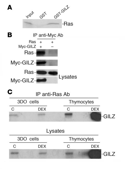
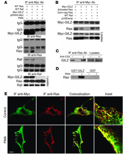
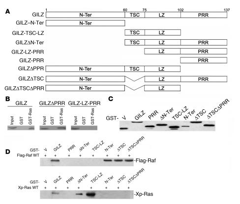
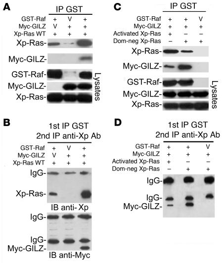
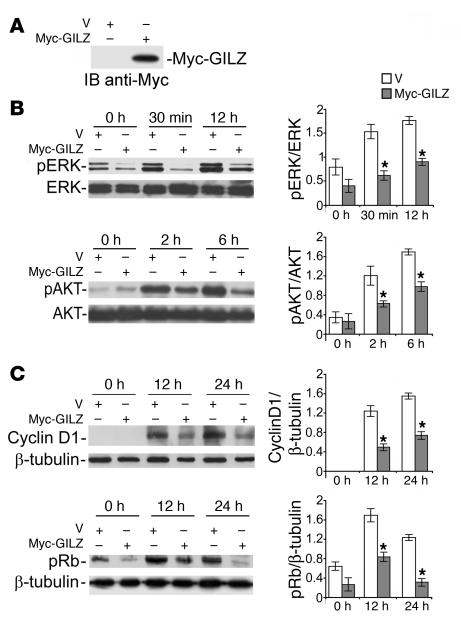
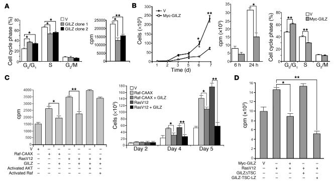
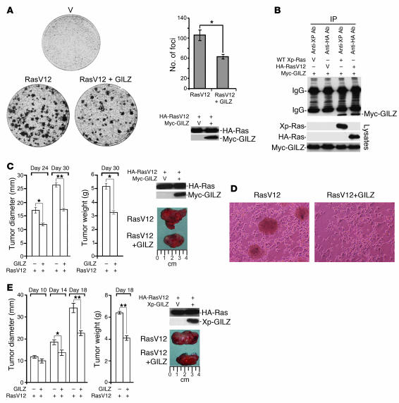
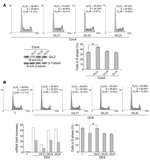
Tsc22d3 coding for glucocorticoid-induced leucine zipper (GILZ) was initially identified as a dexamethasone-responsive gene involved in the control of T lymphocyte activation and apoptosis. However, the physiological role of this molecule and its function in the biological activity of glucocorticoids (GCs) has not been clarified. Here, we demonstrate that GILZ interacts directly with Ras in vitro and in vivo as shown by GILZ and Ras coimmunoprecipitation and colocalization upon PMA activation in primary mouse spleen T lymphocytes and thymus cells. The analysis of GILZ mutants showed that they bound Ras through the tuberous sclerosis complex box (TSC) and, depending on the Ras activation level, formed a trimeric complex with Ras and Raf, which we previously identified as a GILZ binder. As a consequence of these interactions, GILZ diminished the activation of Ras and Raf downstream targets including ERK1/2, AKT/PKB serine/threonine kinase, and retinoblastoma (Rb) phosphorylation and cyclin D1 expression, leading to inhibition of Ras- and Raf-dependent cell proliferation and Ras-induced NIH-3T3 transformation. GILZ silencing resulted in an increase in concanavalin A-induced T cell proliferation and, most notably, inhibition of dexamethasone antiproliferative effects. Together, these findings indicate that GILZ serves as a negative regulator of Ras- and Raf-induced proliferation and is an important mediator of the antiproliferative effect of GCs.


Figures

Figure 1
Figure 1. GILZ interacts with Ras.
(A) The 35S-labeled in vitro transcription and translation product of full-length wild-type H-Ras was incubated with GST or GST-GILZ immobilized on glutathione-sepharose beads. The proteins bound to the resin were eluted, resolved by SDS-PAGE, and visualized by autoradiography. Input indicates 10% volume of the 35S-labeled product used in the pulldown assay. (B) COS-7 cells were cotransfected with pUSEamp-Ras wild-type and Myc-GILZ vectors. Immunoprecipitation was performed with anti-myc Ab, and immunoreactive proteins were revealed with anti-Ras or anti-myc Ab. Whole-cell lysates were loaded to control plasmid expression. (C) 3DO cells and mouse thymocytes were treated for 3 hours with DEX. Cell lysates were immunoprecipitated with anti-Ras Ab and revealed with anti-GILZ Ab. Whole-cell lysates were loaded to control GILZ regulation by DEX. C, control.
Figure 2
Figure 2. GILZ interacts mainly with Ras in the active form.
(A) COS-7 cells were cotransfected with the indicated vectors. After 48 hours, the cells were treated with PMA (100 ng/ml) for 20 minutes. Total cell lysates were subjected to immunoprecipitation with anti-myc Ab and analyzed by immunoblot for the indicated Abs. Whole-cell lysates were loaded to control plasmid expression. (B) COS-7 cells were cotransfected with the indicated vectors, immunoprecipitated, and analyzed by Western blot as described in A. Dom-neg Ras, dominant-negative Ras. (C) Splenic T lymphocytes were activated by cross-linked anti-CD3 Ab for 30 minutes. Cell lysates were immunoprecipitated by anti-Ras Ab and analyzed by immunoblot for anti-GILZ Ab. (D) 3DO cells were treated with PMA (100 ng/ml) for 20 minutes, and pulldown assays were performed by incubating total lysates with GST-GILZ fusion protein or GST alone for 18 hours at 4°C. Immunoblot was analyzed by anti-Ras Ab. (E) Confocal analysis of Ras (red) and GILZ (green) localization in myc-GILZ/pUSEamp-Ras wild-type–cotransfected COS-7 cells treated with PMA for 20 minutes. PMA treatment produced iuxta-membrane accumulation of Ras and an increase in the colocalization with GILZ in the same areas (inset). IF, immunofluorescence. Scale bar, 10 μm. Original magnification, ×5 (top inset); ×6 (bottom inset).
Figure 3
Figure 3. Mapping the Ras-binding domains of GILZ.
(A) Schematic of GILZ deletion constructs. N-Ter, N-terminal. (B) GST-Ras fusion protein was incubated for 18 hours with 35S-labeled in vitro–transcribed and –translated proteins, GILZ, or GILZ mutants. (C) GST-GILZ or GST-GILZ mutants (in pEBG) were transfected in COS-7, and lysates were analyzed by Western blot using anti-GST Ab to control plasmid expression. (D) COS-7 cells were cotransfected with GST-GILZ or GST-GILZ mutants, as described in C, together with either Flag-Raf (top) or Xp-Ras (bottom). GST-GILZ and GST-GILZ mutants were purified by glutathione-sepharose resin, and the copurified Raf and Ras were detected by anti-Flag and anti-Xp Abs, respectively. V, empty vector.
Figure 4
Figure 4. GILZ forms a ternary complex with Ras and Raf.
(A) GST-Raf and wild-type Xp-Ras were transfected in COS-7 cells with or without myc-GILZ. GST-Raf was purified by adsorption to glutathione-sepharose beads, and copurified proteins were revealed with anti-Xp and anti-myc Abs using Western blot. Lysates were loaded to control expression levels. (B) The overexpressed glutathione-purified proteins were subjected to immunoprecipitation with anti-Xp Ab, followed by Western blot with anti-Xp and anti-myc antibodies. (C) GST-Raf and myc-GILZ were transfected in COS-7 cells together with either activated or dominant-negative Xp-Ras. Cell lysates, purified by glutathione-sepharose resin, were eluted by glutathione and analyzed by Western blot with the indicated Abs. (D) The glutathione-purified proteins described in C were immunoprecipitated with anti-Xp Ab and revealed by Western blot with anti-myc Ab.
Figure 5
Figure 5. GILZ inhibits Ras signaling.
NIH-3T3 cells, transfected with myc-GILZ or control vector, were cultured for 12 days with G418, starved for 24 hours (1% FCS), and then stimulated with 10% FCS for the indicated times. Total cell lysates were subjected to Western blot with the indicated Abs. Results of quantitative densitometry analysis are shown at right. (A) GILZ expression evaluated 12 days after transfection. (B) Phosphorylated ERK (pERK) and AKT. (C) Cyclin D1 expression and phosphorylated Rb. After stripping, membranes were reprobed with anti-ERK1/2 or anti-AKT or with anti–β-tubulin Ab. Three independent lysates for each condition were immunoblotted and the data were quantified and expressed as the relative ratio of phosphoprotein to total protein or β-tubulin (mean ± SD). *P < 0.05.
Figure 6
Figure 6. GILZ impairs cell proliferation rate.
(A) Empty vector– or GILZ-transfected 3DO stable clones (clones 1 and 2) were starved for 48 hours and then cultured for 24 hours in the presence of 10% FCS. Proliferation was evaluated by PI staining and cell cycle analysis (left) and [3H]-thymidine uptake assays (right). (B) NIH-3T3 cells were transfected, selected, and controlled for vector expression by Western blot as described in Figure 5, then starved for 24 hours (1% FCS). Growth curves (left) and [3H]-thymidine uptake (center) were quantified following FCS stimulation at the indicated times. Cell cycle analysis of PI-labeled cells (right) was performed 24 hours after FCS stimulation. (C) Inhibitory effect of GILZ on NIH-3T3 cell proliferation, transiently transfected with Raf-CAAX or RasV12 and reversion of GILZ-mediated inhibition by activated AKT or activated Raf. [3H]-thymidine uptake (left) and cell number (right) are shown. (D) Cell proliferation of NIH-3T3 cells transfected with RasV12 and full-length GILZ or GILZ mutant lacking the TSC or expressing only the TSC and LZ regions, as evaluated by [3H]-thymidine uptake. Data represent the average of 3 independent experiments; triplicate samples were counted at each time point. *P < 0.05; **P < 0.01.
Figure 7
Figure 7. GILZ reduces Ras-driven transformation and tumorigenicity.
(A) NIH-3T3 cells were transiently transfected with HA-tagged RasV12 together with either control or myc-GILZ vector. After 14 days, the dishes were stained with 0.1% crystal violet. Quantification of the foci in each dish and the expression of transfected vectors are shown at right. The data represent an average from 3 dishes in 3 independent experiments each. (B) Lysates of NIH-3T3 cells transiently cotransfected with Myc-GILZ and either HA-RasV12 or wild-type Xp-Ras were immunoprecipitated with anti-HA and anti-Xp Abs, respectively. Immunoreactive proteins were revealed with anti-myc Ab. Whole-cell lysates were loaded to control plasmid expression. (C) NIH-3T3 cells were transiently transfected as described above and injected subcutaneously into SCID mice (5 per group). Tumor growth was scored at the indicated times as tumor diameter and tumor weight (left). Plasmid expression was evaluated 2 days after transfection by Western blot (right). (D) NIH-3T3 cells transfected with HA-RasV12 with or without Xp-GILZ were double-selected with antibiotics, and plates were photographed 2 weeks after transfection, at which time the cells were used to assess expression levels by Western blot with the Abs indicated in E. (E) The cells described in D were subcutaneously injected into SCID mice, and tumor growth was scored at the indicated times. After excision, representative tumors were photographed by a digital camera. *P < 0.05; **P < 0.01.
Figure 8
Figure 8. GILZ controls T cell proliferation and is responsible for GC-mediated anti-proliferative activity.
(A) Splenic T lymphocytes were transfected with GILZ1, GILZ2, or GILZs siRNAs and stimulated with ConA (1 μg/ml) for 3 days. Shown are the cell cycle, evaluated by PI assay (DNA content is shown on x axis and number of nuclei is shown on y axis), and GILZ expression, evaluated by Western blot, of a representative experiment. (B) 3DO cells were transfected with GILZ1, GILZ2, or GILZs siRNAs 1 hour before and 3 hours after DEX treatment (100 nM) (DNA content is shown on x axis and number of nuclei is shown on y axis). Shown are GILZ mRNA levels by real-time PCR 6 and 18 hours (white and gray bars, respectively) after silencing and cell cycle analysis by PI assay 18 hours after silencing, from a representative experiment. Histograms in A and B represent the average percent of cells in S phase from 3 independent experiments. *P < 0.05.

Similar articles

Cited by

References

    1. Payne D.N., Adcock I.M. Molecular mechanisms of corticosteroid actions. Paediatr. Respir. Rev. 2001;2:145–150. - PubMed
    1. De Bosscher K., Vanden Berghe W., Haegeman G. Mechanisms of anti-inflammatory action and of immunosuppression by glucocorticoids: negative interference of activated glucocorticoid receptor with transcription factors. J. Neuroimmunol. 2000;109:16–22. - PubMed
    1. Adcock I.M. Glucocorticoid-regulated transcription factors. Pulm. Pharmacol. Ther. 2001;14:211–219. - PubMed
    1. Wikstrom A.C. Glucocorticoid action and novel mechanisms of steroid resistance: role of glucocorticoid receptor-interacting proteins for glucocorticoid responsiveness. J. Endocrinol. 2003;178:331–337. - PubMed
    1. Almawi W.Y., Abou Jaoude M.M., Li X.C. Transcriptional and post-transcriptional mechanisms of glucocorticoid antiproliferative effects. Hematol. Oncol. 2002;20:17–32. - PubMed

Publication types

MeSH terms