. 2007 Aug 15;406(1):97-103.
doi: 10.1042/BJ20070446.

The chondroitin sulfate form of invariant chain trimerizes with conventional invariant chain and these complexes are rapidly transported from the trans-Golgi network to the cell surface

Affiliations

The chondroitin sulfate form of invariant chain trimerizes with conventional invariant chain and these complexes are rapidly transported from the trans-Golgi network to the cell surface

Lynne S Arneson et al. Biochem J. .

Abstract

Targeting of MHCII-invariant chain complexes from the trans-Golgi network to endosomes is mediated by two di-leucine-based signals present in the cytosolic domain of invariant chain. Generation of this endosomal targeting signal is also dependent on multimerization of the invariant chain cytosolic domain sequences, mediated through assembly of invariant chain into homotrimers. A small subset of invariant chain is modified by the addition of chondroitin sulfate and is expressed on the cell surface in association with MHCII. In the present study, we have followed the biosynthetic pathway and route of intracellular transport of this proteoglycan form of invariant chain. We found that the efficiency of chondroitin sulfate modification can be increased by altering the invariant chain amino acid sequence around Ser-201 to the xylosylation consensus sequence. Our results also indicate that, following sulfation, the proteoglycan form is transported rapidly from the trans-Golgi network to the cell surface and is degraded following internalization into an endocytic compartment. Invariant chain-chondroitin sulfate is present in invariant chain trimers that also include conventional non-proteoglycan forms of invariant chain. These data indicate that invariant chain-chondroitin sulfate-containing complexes are transported rapidly from the trans-Golgi network to the cell surface in spite of the presence of an intact endosomal localization signal. Furthermore, these results suggest that invariant chain-chondroitin sulfate may play an important role in the generation of cell-surface pools of invariant chain that can serve as receptors for CD44 and macrophage migration inhibitory factor.


Figures

Figure 1
Figure 1. Sequence of the xylosylation site in murine Ii
The wild-type sequence of Ii (amino acids 196–205) is indicated, along with the sequence of the mutated p31EA construct and the consensus xylosylation sequence [43]. The xylose-accepting residue is in bold (Ser-201), and the mutated amino acids are underlined.
Figure 2
Figure 2. Changing the xylosylation site to conform to the consensus sequence results in increased levels of Ii–CS expression
Ltk− cells were transiently transfected with 0.1, 0.3 or 1.0 μg of cDNA encoding either wildtype p31 or p31EA. The protein expressed by these transfectants was labelled with either [35S]methionine for 10 min or 35SO4 for 30 min. Cell lysate was then immunoprecipitated with P4H5 (anti-Ii) and analysed by SDS/PAGE. The positions of Ii–CS and p31 are indicated on the right, and the positions of the molecular-mass markers are indicated on the left (sizes in kDa).
Figure 3
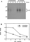
Figure 3. Ii–CS travels rapidly to the cell surface following sulfation in the TGN
(A) Splenocytes isolated from C3H mice were labelled with 35SO4 for 15 min and chased for 0, 15, 30 or 45 min at 37 °C. After each chase, cells were treated with or without chondroitinase ABC. After removal of the chondroitinase by washing, the cells were lysed, immunoprecipitated with P4H5 (anti-Ii) and analysed by SDS/PAGE. Position of molecular-mass markers are indicated on the left (sizes in kDa). (B) Optical densitometry was performed on autoradiographs from two independent experiments, and results are means for cells treated with or without chondroitinase ABC. Similar results were obtained with Ltk− transfectants expressing p31EA (not shown).
Figure 4
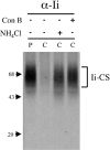
Figure 4. Degradation of Ii–CS is inhibited by lysosomotropic agents
Ltk− cells stably transfected with p31EA were labelled for 1 h with 35SO4 (P) and chased for 1 h (C) without treatment or with 15 mM NH4Cl or 10 μM concanamycin B. Cell lysates were immunoprecipitated with P4H5 (anti-Ii) and analysed by SDS/PAGE. The position of Ii–CS is shown on the right, and molecular-mass markers are indicated on the left (sizes in kDa).
Figure 5
Figure 5. Ii–CS cross-links to form a complex whose molecular mass is consistent with Ii trimerization
Ltk− cells stably transfected with the p31EA construct were labelled with [3H]leucine or 35SO4 for 1.5 h and lysed in the absence (−) or presence (+) of DSP. Lysates were immunoprecipitated with P4H5 (anti-Ii), treated without (A) or with (B) the reducing agent 2-mercaptoethanol to reduce the cross-linker DSP and separated on 5–10% gradient gels. Monomeric, dimeric and trimeric conventional Ii and molecular-mass markers are indicated on the left (sizes in kDa), and Ii–CS complexes are indicated on the right.
Figure 6
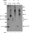
Figure 6. Core Ii–CS is present in complexes whose molecular mass is consistent with trimerization
Ltk− cells stably transfected with the p31EA construct were labelled with [35S]methionine for 2 h and lysed in the absence (−) or presence (+) of the reducible cross-linker DSP. Lysates were immunoprecipitated with P4H5 (anti-Ii) and eluted. Eluates were then either not treated, or absorbed with DEAE to isolate complexes containing Ii–CS. DEAE-bound material was then eluted, treated without (−) or with (+) chondroitinase ABC (Case) and reprecipitated with a combination of conformation-independent antibodies directed at the cytosolic domain of Ii (In-1 and antisera 657 and 658) and analysed by non-reducing SDS/PAGE. The positions of the monomeric, dimeric and trimeric conventional Ii complexes are labelled on the left. The positions of the core-Ii–CS following chondroitinase treatment (core Ii–CS) and the cross-linked complexes containing Ii–CS both before (*) and after (**) chondroitinase treatment are indicated on the right. The position of the high-molecular-mass Ii aggregates (Δ) is indicated on the right. Molecular-mass markers are indicated on the left (sizes in kDa).
Figure 7
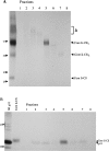
Figure 7. Ii–CS is present in trimers that may contain both CS modified and unmodified Ii
Ltk− cells stably transfected with p31 were labelled with [35S]methionine for 2 h and lysed in the presence of 200 μg/ml DSP. Lysates were absorbed to DEAE and digested with chondroitinase as described in Figure 6. Anti-Ii immunoprecipitates from DEAE-bound and chondroitinase-treated material was eluted in the absence of 2-mercaptoethanol and run on 7% tube gels. The tube gel was then cut into 1 cm pieces (fractions 1–8), protein was eluted overnight at room temperature, unreduced (A) or reduced (B) to break the cross-linking and separated on 5–10% gradient gels. Lane 1 in (A) contains an aliquot of the material loaded on the tube gel (DSP cross-linked, DEAE-absorbed, chondroitinase-digested and immunoprecipitated with anti-Ii). The positions of the high-molecular-mass Ii aggregates (Δ) and the putative trimeric, dimeric and monomeric p31–CS core complexes are indicated on the right. Lanes 1 and 2 in (B) contain control lanes to indicate the position of [35S]methionine-labelled p31 and p31–CS core proteins. Lane 1 is an anti-Ii precipitate from untreated lysates. The sample in lane 2 was first absorbed to DEAE to enrich for Ii–CS, chondroitinase-treated and then precipitated with anti-Ii. The position of p31 and the slower migrating core Ii–CS are indicated on the right. Molecular-mass markers are indicated on the left of both gels (sizes in kDa).

Similar articles

Cited by

References

    1. Hiltbolt E. M., Roche P. A. Trafficking of MHC class II molecules in the late secretory pathway. Curr. Opin. Immunol. 2002;14:30–35. - PubMed
    1. Bakke O., Dobberstein B. MHC class II-associated invariant chain contains a sorting signal for endosomal compartments. Cell. 1990;63:707–716. - PubMed
    1. Lotteau V., Teyton L., Peleraux A., Nilsson T., Karlsson L., Schmid S., Quaranta V., Peterson P. Intracellular transport of class II MHC molecules directed by invariant chain. Nature. 1990;348:600–605. - PubMed
    1. Shachar I., Flavell R. Requirement for invariant chain in B cell maturation and function. Science. 1996;274:106–108. - PubMed
    1. Benlagha K., Park S.-H., Guinamard R., Forestier C., Karlsson L., Chang C.-H., Bendelac A. Mechanisms governing B cell development defects in invariant chain-deficient mice. J. Immunol. 2004;172:2076–2083. - PubMed

Publication types

MeSH terms

Substances