. 2007 Aug 15;406(1):157-65.
doi: 10.1042/BJ20070359.

Phospholipase C beta3 is a key component in the Gbetagamma/PKCeta/PKD-mediated regulation of trans-Golgi network to plasma membrane transport

Affiliations

Phospholipase C beta3 is a key component in the Gbetagamma/PKCeta/PKD-mediated regulation of trans-Golgi network to plasma membrane transport

Alberto M Díaz Añel. Biochem J. .

Abstract

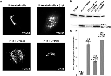
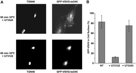
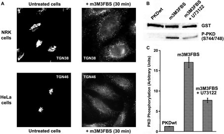
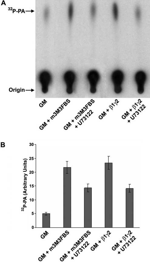
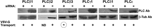
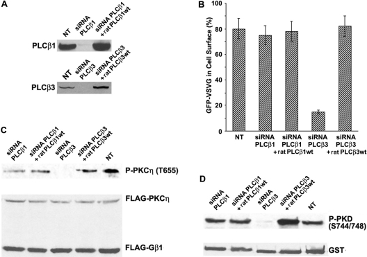
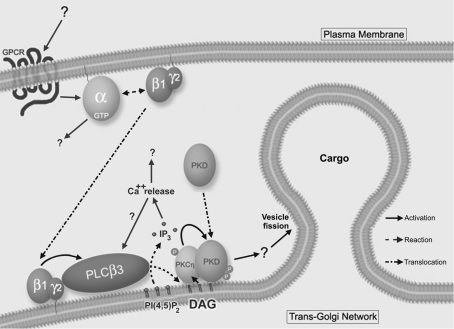
The requirement of DAG (diacylglycerol) to recruit PKD (protein kinase D) to the TGN (trans-Golgi network) for the targeting of transport carriers to the cell surface, has led us to a search for new components involved in this regulatory pathway. Previous findings reveal that the heterotrimeric Gbetagamma (GTP-binding protein betagamma subunits) act as PKD activators, leading to fission of transport vesicles at the TGN. We have recently shown that PKCeta (protein kinase Ceta) functions as an intermediate member in the vesicle generating pathway. DAG is capable of activating this kinase at the TGN, and at the same time is able to recruit PKD to this organelle in order to interact with PKCeta, allowing phosphorylation of PKD's activation loop. The most qualified candidates for the production of DAG at the TGN are PI-PLCs (phosphatidylinositol-specific phospholipases C), since some members of this family can be directly activated by Gbetagamma, utilizing PtdIns(4,5)P2 as a substrate, to produce the second messengers DAG and InsP3. In the present study we show that betagamma-dependent Golgi fragmentation, PKD1 activation and TGN to plasma membrane transport were affected by a specific PI-PLC inhibitor, U73122 [1-(6-{[17-3-methoxyestra-1,3,5(10)-trien-17-yl]amino}hexyl)-1H-pyrrole-2,5-dione]. In addition, a recently described PI-PLC activator, m-3M3FBS [2,4,6-trimethyl-N-(m-3-trifluoromethylphenyl)benzenesulfonamide], induced vesiculation of the Golgi apparatus as well as PKD1 phosphorylation at its activation loop. Finally, using siRNA (small interfering RNA) to block several PI-PLCs, we were able to identify PLCbeta3 as the sole member of this family involved in the regulation of the formation of transport carriers at the TGN. In conclusion, we demonstrate that fission of transport carriers at the TGN is dependent on PI-PLCs, specifically PLCbeta3, which is necessary to activate PKCeta and PKD in that Golgi compartment, via DAG production.


Figures

Figure 1
Figure 1. Inhibitory effect of U73122 on β1γ2-dependent Golgi fragmentation and PKD activation
(A) Purified His–β1γ2 was added to semi-permeabilized NRK cells, previously incubated with U73122 (lower right panel) or U73343 (lower left panel). After 30 min, the cells were treated for immunofluorescence, and Golgi membranes were detected with anti-TGN48 antibodies. Untreated cells were incubated only with (upper right panel) or without (upper left panel) purified His–β1γ2. (B) HeLa cells were co-transfected with FLAG–β1–HA–γ2 and GST–PKD1, and incubated for 16 h in the presence of U73122 or U73343. Cell lysates were prepared and analysed by Western blot to monitor PKD1 activity (activation loop phosphorylation). (C) Densitometry of the experiment described in (B). All the experiments shown in (A) were repeated five times, and values in (C) are the means (±S.D., vertical bars) for four separate experiments.
Figure 2
Figure 2. U73122 blocking effect on TGN to plasma membrane transport
(A) HeLa cells were transfected with the thermosensitive mutant GFP–VSV-G tsO45. After accumulation of this protein in TGN at 20 °C, the cells were incubated at the permissive temperature for 60 min, and then analysed by immunofluorescence to monitor the localization of VSV-G. All the procedure was performed in the presence of U73122 or U73343, except for non-treated cells (NT). (B) Measurement of the experiment described in (A), depicted as a percentage of GFP–VSV-G-expressing cells where this protein has reached the cell surface. Values are the means (±S.D., vertical bars) for three separate experiments.
Figure 3
Figure 3. PI-PLC activator m-3M3FBS induces Golgi fragmentation and PKD1 activation
(A) NRK and HeLa cells were incubated respectively with 25 and 50 μM m-3M3FBS for 30 min. Then, the integrity of TGN structure was analysed by immunofluorescence. (B) Lysed extracts from GST–PKD1-transfected HeLa cells were analysed by Western blot to measure PKD1 activation. In some experiments, cells were incubated with U73122 before and during m-3M3FBS treatment (third lane from left). (C) Densitometry of the experiment described in (B). Values are means (±S.D., vertical bars) for three separate experiments.
Figure 4
Figure 4. β1γ2 and m-3M3FBS induce DAG production at similar levels
(A) Purified rat Golgi membranes (GM) were incubated with purified His–β1γ2 or m-3M3FBS in the presence or absence of U73122. Then, the membrane lipids were extracted and subjected to a DGK assay, as described in the Experimental section, and finally they were analysed by TLC for DAG production, which was measured as quantity of [32P]PA. (B) Densitometry of the experiment described in (A). Values are means (±S.D., vertical bars) for five separate experiments.
Figure 5
Figure 5. Expression of different PI-PLCs' siRNAs, and their effect on VSV-G transport
HeLa cells were transfected with siRNAs in order to block specific PI-PLCs. The resulting extracts prepared 72 h post-transfection were analysed by Western blot for each corresponding PLC (left upper lane for each protein), and then compared with non-transfected cellular extracts (right upper lane). As a control for protein expression, the extracts were monitored for β-tubulin expression (lower lanes). In a parallel experiment, siRNA-treated cells were transfected with GFP–VSV-G tsO45 48 h after siRNA transfection. After 24 h of VSV-G expression, cells were temperature-shifted as described in the Experimental section, and analysed by immunofluorescence to monitor the localization of VSV-G in the cell. Blocking of TGN to plasma membrane transport (circled ‘–’) was considered when less than 20% of GFP–VSV-G reached the cell surface, and normal transport (+) when more than 75% of the VSV-G-expressing cells had this protein localized at the plasma membrane. No data (ND) were collected from PLCβ2-siRNA-transfected cells, since they died 24–36 h post-siRNA transfection. Corresponding molecular masses in kDa are: PLCβ1: 134; PLCβ2: 130; PLCβ3: 132; PLCγ1: 142; PLCδ1: 83; and PLCϵ: 250. All siRNA Western blots and transport experiments were repeated four times.
Figure 6
Figure 6. PLCβ3 siRNA blocks VSV-G transport and PKCη/PKD activation
(A) Western blots showing the expression of PLCβ1 and PLCβ3 in normal cells (NT), siRNA-transfected cells, and siRNA-treated cells where the corresponding rat wild-type protein (ratPLCβ1 or 3 wt) was overexpressed for 24 h before cell lysate preparation. (B) Measurement of VSV-G transport in cells undergoing the same experimental conditions described in (A), shown as a percentage of GFP–VSV-G-expressing cells where this protein has reached the cell surface. Values are means (±S.D., vertical bars) for three separate experiments. (C) Measurement of PKCη activation (Phospho-PKCη Thr655) in extracts proceeding from the cells described in (A). (D) Same as (C) but for PKD1 activation (Phospho-PKD1 Ser744/Ser748). In order to determine activity of protein kinases in (C, D), cells were co-transfected with FLAG–β1–HA–γ2, FLAG–PKCη and GST–PKD1. All the experiments shown in (A, C, D) were repeated four times.
Figure 7
Figure 7. Model for TGN to plasma membrane transport regulation
We propose that a still undiscovered signal is generated at the cell surface via GPCR. This causes the activation of a G-protein, allowing βγ subunits to translocate to the TGN, where they are able to activate PLCβ3 by binding to its PH domain. The activation of this phospholipase leads to the formation of DAG, which has two roles at the TGN: to directly activate PKCη, and to allow PKD1 to translocate to this Golgi compartment by direct binding to its C1a domain. Then, PKCη binds to the PH domain of PKD, which enables its phosphorylation by the former at its activation loop. Finally, PKD1 activation induces cargo-filled vesicle fission at the TGN by phosphorylation of still unknown downstream effectors.

Similar articles

Cited by

References

    1. Takizawa P. A., Yucel J. K., Veit B., Faulkner D. J., Deerinck T., Soto G., Ellisman M., Malhotra V. Complete vesiculation of Golgi membranes and inhibition of protein transport by a novel sea sponge metabolite, ilimaquinone. Cell. 1993;73:1079–1090. - PubMed
    1. Jamora C., Takizawa P. A., Zaarour R. F., Denesvre C., Faulkner D. J., Malhotra V. Regulation of Golgi structure through heterotrimeric G proteins. Cell. 1997;91:617–626. - PubMed
    1. Jamora C., Yamanouye N., Van Lint J., Laudenslager J., Vandenheede J. R., Faulkner D. J., Malhotra V. Gbetagamma-mediated regulation of Golgi organization is through the direct activation of protein kinase D. Cell. 1999;98:59–68. - PubMed
    1. Díaz Añel A. M., Malhotra V. PKCeta is required for beta1gamma2/beta3gamma2- and PKD-mediated transport to the cell surface and the organization of the Golgi apparatus. J. Cell Biol. 2005;169:83–91. - PMC - PubMed
    1. Liljedahl M., Maeda Y., Colanzi A., Ayala I., Van Lint J., Malhotra V. Protein kinase D regulates the fission of cell surface destined transport carriers from the trans-Golgi network. Cell. 2001;104:409–420. - PubMed

Publication types

MeSH terms

Substances