Progressive supranuclear palsy: pathology and genetics
- PMID: 17493041
- PMCID: PMC8095545
- DOI: 10.1111/j.1750-3639.2007.00054.x
Progressive supranuclear palsy: pathology and genetics
Abstract
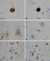
Progressive supranuclear palsy (PSP) is an atypical Parkinsonian disorder associated with progressive axial rigidity, vertical gaze palsy, dysarthria and dysphagia. Neuropathologically, the subthalamic nucleus and brainstem, especially the midbrain tectum and the superior cerebellar peduncle, show atrophy. The substantia nigra shows loss of pigment corresponding to nigrostriatal dopaminergic degeneration. Microscopic findings include neuronal loss, gliosis and neurofibrillary tangles in basal ganglia, diencephalon and brainstem. Characteristic tau pathology is also found in glia. The major genetic risk factor for sporadic PSP is a common variant in the gene encoding microtubule-associated protein tau (MAPT) and recent studies have suggested that this may result in the altered expression of specific tau protein isoforms. Imaging studies suggest that there may be sensitive and specific means to differentiate PSP from other parkinsonian disorders, but identification of a diagnostic biomarker is still elusive.
Figures


References
-
- Andreadis A, Brown WM, Kosik KS (1992) Structure and novel exons of the human tau gene. Biochemistry 31:10626–10633. - PubMed
-
- Arai H, Morikawa Y, Higuchi M, Matsui T, Clark CM, Miura M, Machida N, Lee VM, Trojanowski JQ, Sasaki H (1997) Cerebrospinal fluid tau levels in neurodegenerative diseases with distinct tau‐related pathology. Biochem Biophys Res Commun 236:262–264. - PubMed
-
- Arima K, Nakamura M, Sunohara N, Ogawa M, Anno M, Izumiyama Y, Hirai S, Ikeda K (1997) Ultrastructural characterization of the tau‐immunoreactive tubules in the oligodendroglial perikarya and their inner loop processes in progressive supranuclear palsy. Acta Neuropathol (Berl) 93:558–566. - PubMed
-
- Baker M, Litvan I, Houlden H, Adamson J, Dickson D, Perez‐Tur J, Hardy J, Lynch T, Bigio E, Hutton M (1999) Association of an extended haplotype in the tau gene with progressive supranuclear palsy. Hum Mol Genet 8:711–715. - PubMed
-
- Baker M, Mackenzie IR, Pickering‐Brown SM, Gass J, Rademakers R, Lindholm C, Snowden J, Adamson J, Sadovnick AD, Rollinson S, Cannon A, Dwosh E, Neary D, Melquist S, Richardson A, Dickson D, Berger Z, Eriksen J, Robinson T, Zehr C, Dickey CA, Crook R, McGowan E, Mann D, Boeve B, Feldman H, Hutton M (2006) Mutations in progranulin cause tau‐negative frontotemporal dementia linked to chromosome 17. Nature 442: 916–919. - PubMed
Publication types
MeSH terms
Substances
Grants and funding
- P01-AG17216/AG/NIA NIH HHS/United States
- P50 NS040256/NS/NINDS NIH HHS/United States
- P50 AG016574/AG/NIA NIH HHS/United States
- P01-AG14449/AG/NIA NIH HHS/United States
- R01 AG015866/AG/NIA NIH HHS/United States
- R01 ES013941/ES/NIEHS NIH HHS/United States
- P50-AG16574/AG/NIA NIH HHS/United States
- P01 AG014449/AG/NIA NIH HHS/United States
- P50-NS40256/NS/NINDS NIH HHS/United States
- R01-ES02941/ES/NIEHS NIH HHS/United States
- P50 AG025711/AG/NIA NIH HHS/United States
- P01 AG017216/AG/NIA NIH HHS/United States
- P01 NS040256/NS/NINDS NIH HHS/United States
- P50-AG25711/AG/NIA NIH HHS/United States
- P01 AG003949/AG/NIA NIH HHS/United States
- R01-AG15866/AG/NIA NIH HHS/United States
- P01-AG03949/AG/NIA NIH HHS/United States
LinkOut - more resources
Full Text Sources
Medical
Miscellaneous

