

Review
doi: 10.1111/j.1750-3639.2007.00053.x.
Structural principles of tau and the paired helical filaments of Alzheimer's disease
Affiliations
- PMID: 17493042
- PMCID: PMC8095506
- DOI: 10.1111/j.1750-3639.2007.00053.x
Item in Clipboard
Review
Structural principles of tau and the paired helical filaments of Alzheimer's disease
Brain Pathol.
2007 Jan.
Abstract
Tau, a major microtubule-associated protein in brain, forms abnormal fibers in Alzheimer's disease and several other neurodegenerative diseases. Tau is highly soluble and adopts a natively unfolded structure in solution. In the paired helical filaments of Alzheimer's disease, small segments of tau adopt a beta-conformation and interact with other tau molecules. In the filament core, the microtubule-binding repeat region of tau has a cross-beta structure, while the rest of the protein retains its largely unfolded structure and gives rise to the fuzzy coat of the filaments.
Figures
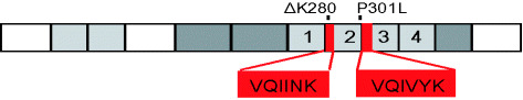
Diagram of tau441, the longest isoform in human brain. The four repeats of 31 or 32 residues each are numbered. The inserts near the N‐terminus and the second repeat can be alternatively spliced, giving rise to six isoforms. The N‐terminal domain up to ∼G120 has an acidic character, the other domains are basic. The left half (residues 1 to ∼200) represents the “projection domain”, the right half the “microtubule assembly domain”. The repeats constitute the core of the microtubule‐binding domain, as well as the core of the paired helical filaments. Two hexapeptide motifs at the beginning of R2 and R3 promote paired helical filament (PHF) aggregation by inducing β‐structure. ΔK280 and P301L are two FTDP‐17 (frontotemporal dementia and parkinsonism linked to chromosome 17) mutations that strongly enhance the rate of PHF aggregation by increasing the propensity for β‐structure.

Model of microtubule protofilament with bound kinesin and tau. The protofilament consists of alternating subunits of α‐ and β‐tubulin (∼450 residues each) arranged in a polar fashion. The head domain of kinesin, a microtubule‐dependent motor protein (∼350 residues), has the compact folding typical of most cytoplasmic proteins. By contrast, tau is natively unfolded, its structure is unknown in detail and modeled here as a random chain. Note that tau occupies a much larger volume than kinesin or tubulin.
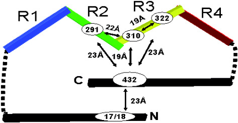
Model of the conformation of tau in solution deduced by fluorescence resonance energy transfer. The molecule shows a paperclip‐like fold which brings the N‐ and C‐terminal ends into the vicinity of the repeat domain. Similar folded conformations are recognized by several antibodies specific for abnormal tau from Alzheimer’s disease brain (eg, Alz‐50, MC1, TG3). Approximate distances between labeled residues are indicated.
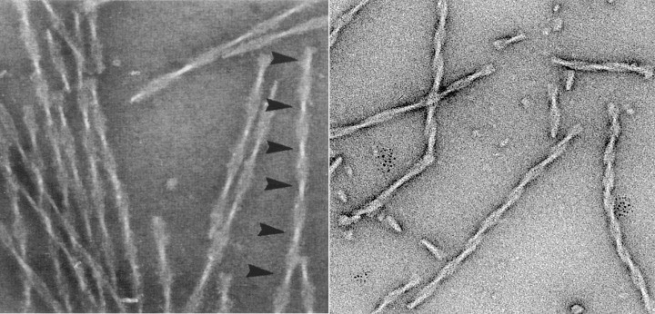
Electron micrographs of paired helical filaments isolated from Alzheimer’s disease brain (left) or assembled in vitro from recombinant tau (repeat domain with pro‐aggregation mutation ΔK280). Note the typical twisted appearance with crossover repeats of ∼80 nm (arrowheads).
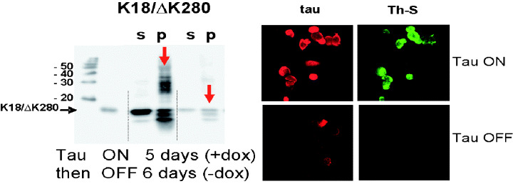
Inducible expression of tau in N2a cells. The panel on the left illustrates that the expression of tau can be switched on by doxycyclin, followed by aggregation (note the high‐molecular weight smear in the gel; s = soluble tau, p = aggregated tau). Expression of tau and aggregates can be reversed by removal of doxycyclin. The panel on the right shows expression of tau (red) and formation of aggregates, as seen by thioflavin S staining (green).
References
-
- Alzheimer A (1907) Über eine eigenartige Erkrankung der Hirnrinde. Allg Z Psychia 64:146–148.
-
- Andorfer C, Kress Y, Espinoza M, De Silva R, Tucker KL, Barde YA, Duff K, Davies P (2003) Hyperphosphorylation and aggregation of tau in mice expressing normal human tau isoforms. J Neurochem 86:582–590. - PubMed
-
- Andreadis A (2005) Tau gene alternative splicing: expression patterns, regulation and modulation of function in normal brain and neurodegenerative diseases. Biochim Biophys Acta 1739:91–103. - PubMed
-
- Arai T, Guo JP, McGeer PL (2005) Proteolysis of non‐phosphorylated and phosphorylated tau by thrombin. J Biol Chem 280:5145–5153. - PubMed
Publication types
MeSH terms
Substances
LinkOut - more resources
Full Text Sources
Other Literature Sources
Medical
