Review
. 2007 Feb;17(1):117-36.
doi: 10.1016/j.nic.2007.01.003.

Cutting-edge imaging of the spine

Affiliations
Review

Cutting-edge imaging of the spine

A Talia Vertinsky et al. Neuroimaging Clin N Am. 2007 Feb.

Abstract

Damage to the spinal cord may be caused by a wide range of pathologies and generally results in profound functional disability. A reliable diagnostic workup of the spine is very important because even relatively small lesions in this part of the central nervous system can have a profound clinical impact. MR imaging has become the method of choice for the detection and diagnosis of many spine disorders. Various innovative MR imaging methods have been developed to improve neuroimaging, including better pulse sequences and new MR contrast parameters. These new "cutting-edge" technologies have the potential to impact profoundly the ease and confidence of spinal disease interpretation and offer a more efficient diagnostic workup of patients suffering from spinal disease.


Figures
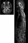
Figure 1
Figure 1
Sagittal T2W FSE of the entire spine and axial T2W FSE through the skull base in a patient with neurofibromatosis type 2. Bilateral masses in cerebello-pontine angles (arrowheads) as well as throughout the spine (arrows) are clearly visible. New multicoil technology affords seemless imaging of the entire CNS without repositioning the patient. (images courtesy Drs. Krueger and Mohr, Siemens Medical Systems, Erlangen, Germany).
Figure 2
Figure 2
Axial views from a modified 3D balanced SSFP (COSMIC) scan of the cervical spine (A) and sagittal reformats (B). The interface between CSF and cord is well identified with superb visualization of nerve roots. Gray and white matter conspicuity is improved over T2W FSE and GRE sequences.
Figure 3
Figure 3
3T Imaging. A. Sagittal T2w (left) and T1w (right) images demonstrate relatively normal spine with mild degenerative changes of the L5-S1 disk (arrow). B. Sag T2w image of the lumbar spine demonstrates hemangioma at L2 (arrowhead) and a mildly protruded disc at L5-S1 (arrow) on T2w scans. In both patients the better resolution afforded by the higher SNR at 3T allows for better delineation of the conus, anatomy of the vertebral bodies and disks.
Figure 4
Figure 4
Axial MERGE image of the cervical spine demonstrates excellent gray/white contrast in the spinal cord as well as good contrast between CSF and the cord (A). The good SNR and contrast helps to demonstrate nerve roots extremely well. MERGE acquires multiple echoes during an oscillatory gradient echo readout (B), which become increasingly T2* weighted (C). The echoes are combined in a way that the early echoes provide increased SNR whilst the later improve contrast.
Figure 5
Figure 5
Sagittal T2w FSE with conventional Fourier encoding (left) and driven equilibrium T2w FSE (FR-FSE) with PROPELLER readout (right). Conventional Fourier encoding is sensitive to pulsation and motion. Such artifacts demonstrate multiple ghosts along the phase encode direction (left, see insert). Since PROPELLER excessively oversamples the center of k-space with each PROPELLER blade, the pulsatile and motion distortions are essentially averaged out (right, see insert). (images courtesy Dr. A. Gaddipati, GE Healthcare).
Figure 6
Figure 6
Positional device (A-D) to A/P (B) or L/R (C) flex and rotate (D) a patient's head and keep it in the desired position for scanning. By performing scans in different head positions, such as anterior flexion (E) or posterior flexion (F) bulging disks (arrows) can be detected that may appear as normal in a neutral position of the neck. Such positional devices allow for an optimal assessment of spinal canal stenosis and neuroforamenal narrowing. This improves correlation between imaging findings and clinical symptoms as patients may experience pain only in certain positions. (images courtesy Drs. Krueger and Mohr, Siemens Medical Systems, Erlangen, Germany).
Figure 7
Figure 7
A. Sagittal CT reformation of the cervical spine in a patient with C4-5 anterior cervical disk fusion. B. The new volumetric FSE sequences (FSE-XETA, VISTA, SPACE) afford much smaller voxel sizes and thus less intravoxel dephasing, improving MR imaging in the presence of metal which is typically problematic because of the field perturbations created by the metal. That in combination with short echo spacing and excessive RF refocusing dramatically reduces distortions in the spine even in the presence of surgical hardware. (images courtesy Dr. Ripart, Siemens Medical Systems, Erlangen, Germany, and Dr. Ricolfi, CHU Dijon, France).
Figure 8
Figure 8
Flip angle sweep during FSE readout. With high flip angles the signal in FSE readouts is primarily determined by the primary echoes, whilst with lower flip angles the signal becomes increasingly dominated by higher order echoes and stimulated echoes. In order to maximize signal for long echo train lengths the flip angle of the refocusing pulses are continuously ramped down almost 50deg and slightly raised when the center of k-space is acquired to optimize contrast and SNR.
Figure 9
Figure 9
Axial view of a volumetric proton-density weighted FSE scan (VISTA) provides excellent gray/white matter contrast (images courtesy Dr. Hoogenraad, Philips Medical Systems, Eindhoven, Netherlands).
Figure 10
Figure 10
Axial T2w FSE images with spectral fat saturation (A) and short-tau inversion recovery (STIR) (B) in a patient with intrapedicular screws for posterior fusion (image below the screws). The geometric distortions from the titanium screws are clearly apparent on spectral fat saturation as increased signal in perivertebral soft tissues as well as in neural foramina. The field perturbations induced by the screws impair chemical fat saturation and lead to difficulties separating between fat and edema or fluid collections, which is significantly reduced on STIR.
Figure 11
Figure 11
Midline sagittal and parasagittal postcontrast T1W fat-saturated SE images (A) and corresponding sagittal and parasagittal postcontrast T1W IDEAL-FSE water images (B) in a patient with neurofibromatosis type 1 and spinal hardware. Numerous enhancing lesions are seen within the neural foramina (large arrows), abutting the spinal cord, and in the paraspinal soft tissues. The lesion adjacent to the spinal cord is better visualized in the IDEAL image (B) compared to the fat-saturated image (A), where failed fat saturation from severe B0 inhomogeneities from metallic hardware degrades signal in the spinal canal near this mass. Large areas of failed fat saturation (small arrows) show uniform suppression of fat in the IDEAL water images. (images courtesy Dr. Reeder, University of Wisconsin).
Figure 12
Figure 12
70 year-old male who presented with progressive myelopathy. A. Sag T2 FR FSE images of the thoracic spine demonstrate edema within the thoracic cord. Multiple intradural serpiginous flow voids are seen consistent with enlarged vascular channels. B. Contrast enhanced MRA with contrast bolus timed for maximal enhancement of the aortic arch was performed to evaluate for suspected dural AV fistula. MIP reformats show the feeding artery arising from the left T8 intercostal artery (arrow) (Aorta is indicated by a star). C. Digital subtraction spinal angiography confirmed the MRA findings (arrow) and embolization of the dural AV Fistula was subsequently performed.
Figure 13
Figure 13
Sagittal T2w FSE images of the spine (A-C) demonstrate two non specific extramedullary/intradural masses requiring a differential diagnosis of several neoplastic etiologies. Sagittal and axial diffusion weighted images (D and E) show these masses to have significantly reduced diffusion thereby making a diagnosis of epidermoids. (images courtesy Dr. M. M. Thurnher, Medical University Vienna, Vienna, Austria).
Figure 14
Figure 14
Functional MRI (fMRI) in the cervical spinal cord during performance of nociceptive pain stimulation on the right upper extremity using a Peltier element to generate local heating. Clearly, activation increases with increased temperature of the Peltier element. To isolate BOLD effect extra caution has to be exercised to minimize the influence from cord pulsation and respiratory artifacts. (images courtesy Drs. Mackey and Glover, Stanford University, Stanford, CA).

Similar articles

Cited by

References

    1. Ghanem N, Uhl M, Brink I, Schafer O, Kelly T, Moser E, Langer M. Diagnostic value of MRI in comparison to scintigraphy, PET, MS-CT and PET/CT for the detection of metastases to bone. European Journal of Radiology. 2005;55:41–55. - PubMed
    1. Roemer PB, Edelstein WA, Hayes CE, Souza SP, Mueller OM. The NMR phased array. Magn Reson Med. 1990 Nov;16(2):192–225. - PubMed
    1. Pruessmann KP, Weiger M, Scheidegger MB, Boesiger P. SENSE: sensitivity encoding for fast MRI. Magn Reson Med. 1999 Nov;42(5):952–62. - PubMed
    1. Griswold MA, Jakob PM, Heidemann RM, Nittka M, Jellus V, Wang J, Kiefer B, Haase A. Generalized autocalibrating partially parallel acquisitions (GRAPPA) Magn Reson Med. 2002 Jun;47(6):1202–10. - PubMed
    1. Hollingworth W, Todd CJ, Bell MI, Arafat Q, Girling S, Karia KR, Dixon AK. The Diagnosis and Therapeutic Impact of MRI: an Observational Multi-centre Study. Clinical Radiology. 2000;55:825–831. - PubMed

MeSH terms