. 2007 Jun 15;306(2):685-702.
doi: 10.1016/j.ydbio.2007.04.006. Epub 2007 Apr 12.

distal antenna and distal antenna-related function in the retinal determination network during eye development in Drosophila

Affiliations

distal antenna and distal antenna-related function in the retinal determination network during eye development in Drosophila

Jennifer Curtiss et al. Dev Biol. .

Abstract

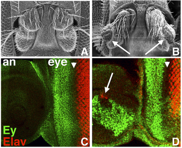
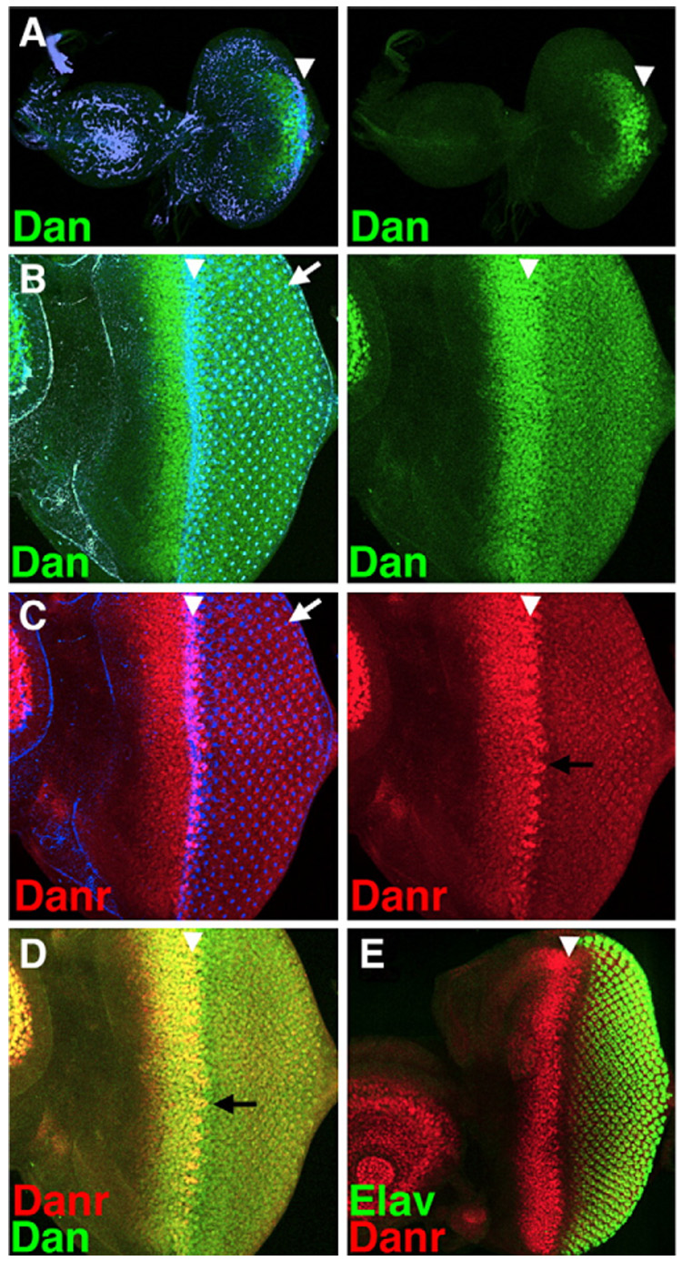
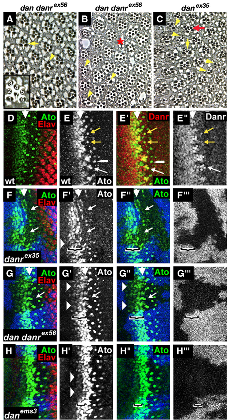
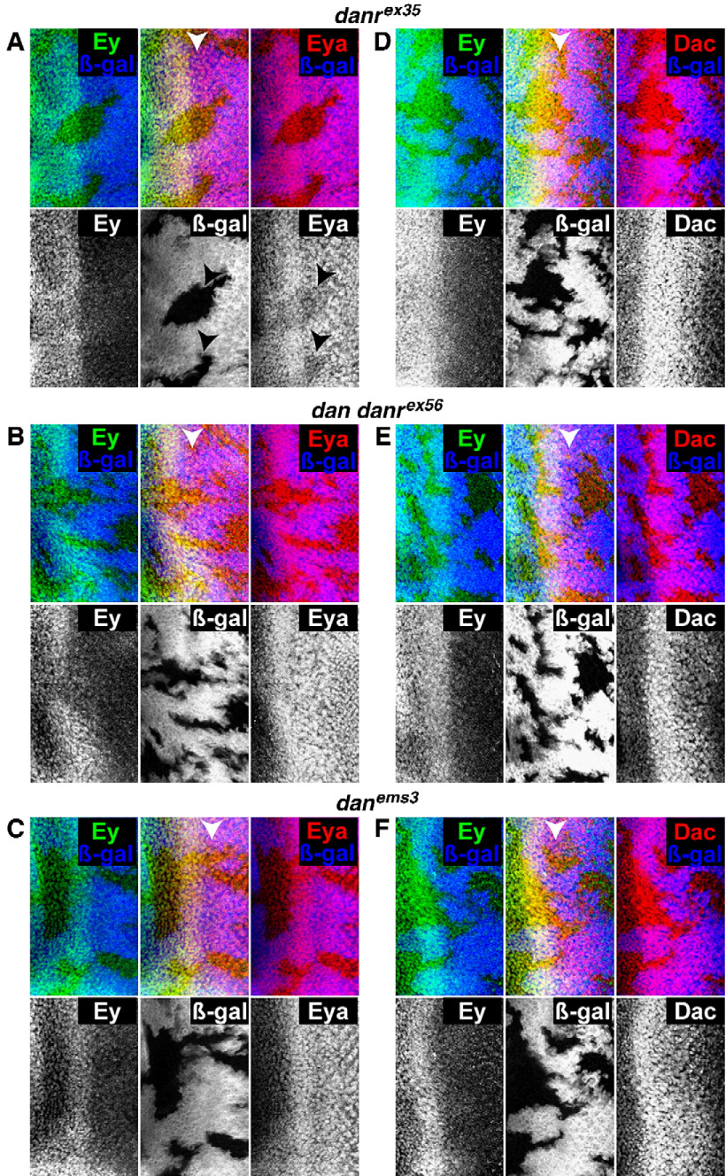
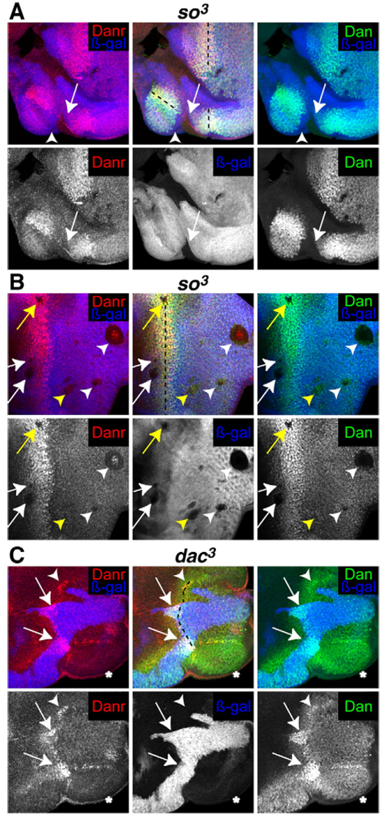
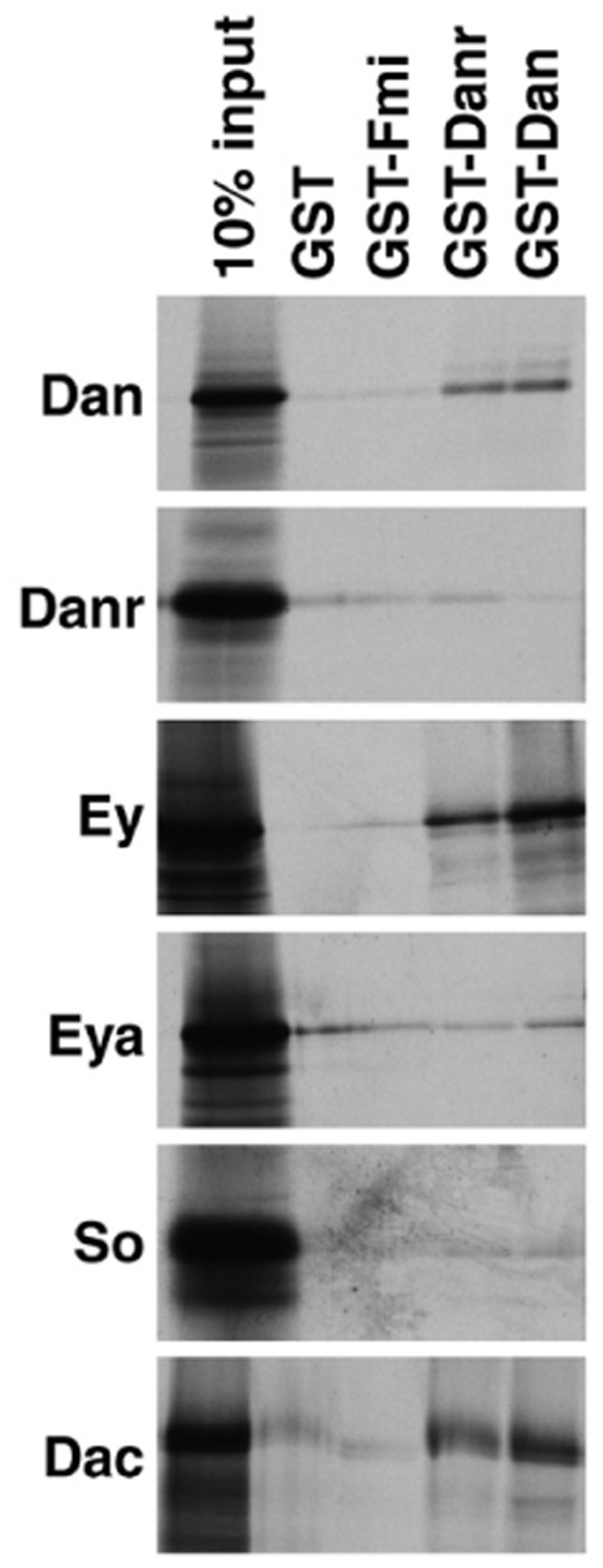
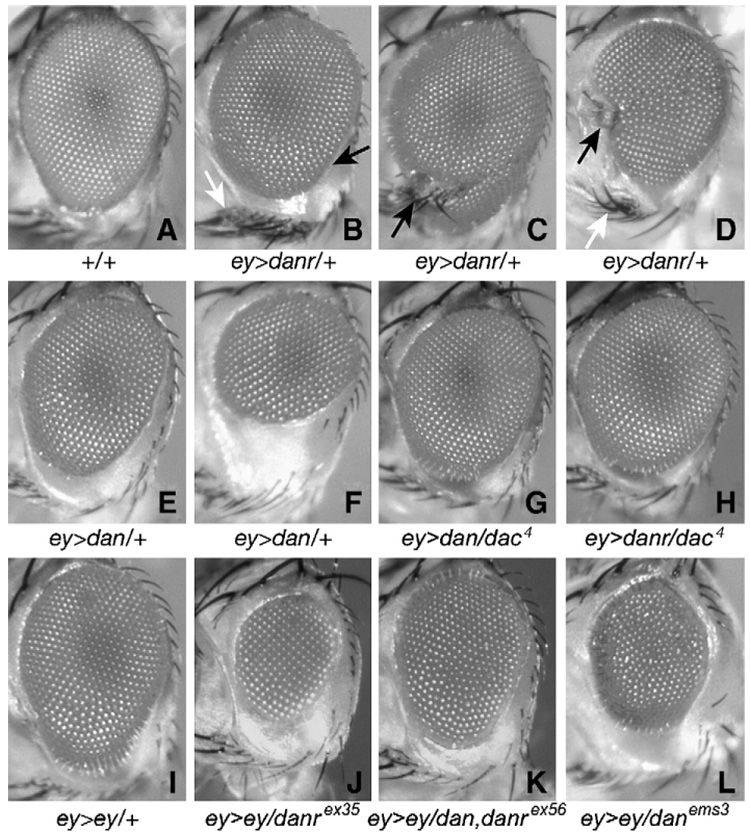
Drosophila eye specification occurs through the activity of the retinal determination (RD) network, which includes the Eyeless (Ey), Eyes absent (Eya), Sine oculis (So) and Dachshund (Dac) transcription factors. Based on their abilities to transform antennal precursors towards an eye fate, the distal antenna (dan) and distal antenna-related (danr) genes encode two new RD factors. Dan and Danr are probable transcription factors localized in nuclei of eye precursors and differentiating eye tissue. Loss-of-function single and double dan/danr mutants have small, rough eyes, indicating a requirement for wild-type eye development. In addition, dan and danr participate in the transcriptional hierarchy that controls expression of RD genes, and Dan and Danr interact physically and genetically with Ey and Dac. Eye specification culminates in differentiation of ommatidia through the activities of the proneural gene atonal (ato) in the founding R8 photoreceptor and Egfr signaling in additional photoreceptors. Danr expression overlaps with Ato during R8 specification, and Dan and Danr regulate Ato expression and are required for normal R8 induction and differentiation. These data demonstrate a role for Dan and Danr in eye development and provide a link between eye specification and differentiation.


Figures

Fig. 1.
Fig. 1.
Misexpression of dan or danr during antennal development leads to ectopic eye formation. (A, B) Frontal view of wild-type (A) and Dll>EPg-dan head (B) showing antennae; each antenna in (B) contains ectopic eye tissue (arrows). (C) Wild-type eye–antenna disc. Ey (green) is expressed only in the eye portion of the disc (eye) anterior to the MF (arrowhead); Elav (red) marks developing photoreceptors posterior to the MF. (D) In Dll>EPg-dan discs, in addition to normal patterns in the eye region, Ey (green) is expressed in a large patch in the antennal portion of the disc (an), and Elav (red) is expressed in ectopic photoreceptor precursors (arrow). Anterior is to the left and dorsal up in panels C and D here, as well as in all panels of all subsequent figures.
Fig. 2.
Fig. 2.
Dan and Danr are expressed during eye and antennal development. (A–C) Left panels show an overlay of Dan (green) or Danr (red) expression with phalloidin staining (blue), which highlights the changes in the actin cytoskeleton that occur within the MF (white arrowhead), as well as the center of each developing photoreceptor cluster (white arrow). Right panels show Dan or Danr expression alone. (A) Dan expression (green) is observed in and around the MF as it initiates at the posterior margin early in the third larval instar. (B–D) All panels show the same disc. In a mid 3rd instar disc, where the MF has progressed a significant distance, Dan (green) is expressed at high levels in all cells anterior to and within the MF and at lower levels in all photoreceptor cells posterior to the MF. Danr (red) is expressed in a similar pattern to Dan anterior to the MF. Its levels drop within and posterior to the MF except in regularly spaced groups of cells within the MF (black arrows). (E) Lower magnification view of an eye–antenna disc showing part of Danr expression in the antenna; also showing the relationship between Danr expression anterior to and within the MF, and Elav expression (green) in the developing photoreceptor cells.
Fig. 3.
Fig. 3.
Loss-of-function mutants for dan/danr have small, rough eyes. (A) Close-up of the MF from a wild-type eye–antenna disc. (B–D) Discs containing loss-of-function dan/danr clones. Dan (green), Danr (red) and clonal marker (blue) are shown. Clones are marked by the absence of blue staining (see Materials and methods), except in panel B″, where the clonal marker has been removed for clarity. Overlay between red and blue, green and blue, and red and green appears pink, turquoise and yellow, respectively. (B) danrex35 clones do not express Danr, but express higher levels of Dan. (C) dan danrex56 clones do not express either Danr or Dan. (D) danems3 clones express normal levels of Dan; Danr expression is lost from clones anterior to the MF (arrows). (E) SEM picture of a wild-type eye. Eyes from eyFLP;FRT82, arm-lacZ,M/FRT82,danrex35 (F) or eyFLP;FRT82,arm-lacZ,M/FRT82,dan danrex56 (G) individuals. Most of both eyes consist of homozygous mutant tissue (see Materials and methods). danrex35 and dan danrex56 eyes are small and rough. (H) Eye from a danems3 homozygote is slightly small.
Fig. 4.
Fig. 4.
danrex35 and dan danrex56 homozygous eye tissues show defects in ommatidial spacing and photoreceptor recruitment. (A–C) Cross sections of eyes from eyFLP;FRT82,arm-lacZ,M/FRT82,dan danrex56 or eyFLP;FRT82,arm-lacZ,M/FRT82,danrex35 flies. Inset: an enlarged wild-type ommatidium showing the positions of the R1–R7 photoreceptors. The internal structure of dan/danr adult mutant eye tissue is variable, ranging from most ommatidia appearing normal (A) to eyes with more frequent defects (B or C) including a loss of photoreceptors (yellow arrowheads), extra small-rhabdomere photoreceptors (C—yellow vertical arrow), defects in rotation (C—red arrow) or gaps in the ommatidial arrangement (B—red asterisk). (D–G) Third instar eye discs. MF is marked by a white arrowhead. (D) Wild-type eye disc double labeled for Elav (red) and Ato (green). (E) Wild-type eye disc double labeled for Ato (green) and Danr (red). Ato and strong levels of Danr expression co-localize in the intermediate groups, in the R8 equivalence group (white arrow) and for a brief period in newly emerged R8 cells (yellow arrows). Shortly afterwards, the strong levels of Danr expression in R8 fade (white long arrowhead). (F–H) Loss-of-function dan/danr clones marked by absence of the blue staining. (F, G) In danrex35 and dan danrex56 mutant clones the stripe of Ato expression (green) anterior to the MF is reduced (horizontal arrowheads), and the number and spacing of the Ato positive R8 cells are altered (arrows). (H) In danems3 mutant clones a narrower stripe of cells ahead of the MF shows a reduction in Ato expression (horizontal arrowheads). The brackets in panels F–H delineate the width of the region ahead of the MF in which Ato expression is reduced in the clones.
Fig. 5.
Fig. 5.
dan/danr and the regulation of RD factor expression. (A–C) Clones marked by the absence of the β-galactosidase (blue) and stained for Ey (green) and Eya (red). (D–F) Clones marked by the absence of the β-galactosidase (blue) and stained for Ey (green) and Dac (red). (A,D) eyFLP; FRT82 arm-lacZ/FRT82 danrex35 discs. Eya is expressed at reduced levels in danrex35 mutant clones (arrows—A), but Ey (A, D) and Dac (D) are expressed normally. (B, E) eyFLP; FRT82 arm-lacZ/FRT82 dan danrex56 discs. (C, F) eyFLP; FRT82 arm-lacZ/FRT82 danems3 discs. Eya (B, C) and Ey (B, C, E, F) and Dac (E, F) are expressed at normal levels in both dan danrex56 and danems3 mutant clones.
Fig. 6.
Fig. 6.
(A, B) Discs from eyFLP; FRT42 arm-lacZ/FRT42 so³. (C) Discs from eyFLP; FRT40 arm-lacZ/FRT40 dac³. (A–C) Null so³ or dac³ clones are marked by the absence of β-galactosidase (blue) and stained with anti-Dan (green) and anti-Danr (red). The position of the MF is marked by a black dotted line. (A) Neither Dan nor Danr are expressed in so³ clones that touch the margin of the disc (arrows), but this effect is nonautonomous (arrowhead). (B) Neither Dan nor Danr are expressed in so³ clones anterior or within the MF (white and yellow arrows), or in large clones posterior to the MF (white arrowheads). Other clones posterior to the MF express Dan and Danr (yellow arrowheads). A single row of cells inside the border of so³ clones located within the MF (yellow arrow) or large clones posterior to the MF (white arrowheads) express low levels of Dan and Danr. (C) Danr is not expressed, and Dan is expressed at low levels in dac³ clones that touch the margin of the disc (asterisk). Dan and Danr are expressed at highest levels in dac+ tissue (arrows) and are expressed at intermediate levels in dac³ tissue through which an MF is progressing (arrowhead).
Fig. 7.
Fig. 7.
Dan and Danr interact physically in vitro with themselves and with Ey and Dac. Aliquots of in vitro-translated [35S]methionine-labeled Dan, Danr, Ey, Eya, So or Dac were incubated with glutathione-agarose beads containing bound GST, GST fused to an unrelated protein (GST-Fmi), GST-Dan and GST-Danr. Bound proteins were fractionated by SDS–PAGE and visualized by autoradiography. 10% of the input is loaded for each in vitro-translated protein for reference. Both GST-Danr and GST-Dan interact specifically with in vitro-translated Dan, Ey and Dac. For extension of this analysis by yeast two-hybrid data see Table 2.
Fig. 8.
Fig. 8.
(A–F) Misexpression of dan or danr interferes with eye development. (A) Eye from an Oregon R fly. (B–D) Eyes from ey-Gal4 UAS-danr/+ flies. (E, F) Eyes from ey-Gal4 UAS-dan/+ flies. Eyes from ey>dan or ey>danr flies are small and rough, and ey>danr also causes patterning defects. (G–L) dan and danr can interact in vivo with ey and dac. Eyes from dac/+;ey-Gal, UAS-dan/+ (G) and dac/+;ey-Gal4 UAS-danr/+ (H) flies are larger and less rough than their ey-Gal4 UAS-dan/+ and ey-Gal4 UAS-danr/+ counterparts. (I) Eyes from ey-Gal4 UAS-ey/+ adults are small and rough. Eyes from ey-Gal4 UAS-ey/danrex35 (J), ey-Gal4 UAS-ey/dan danrex56 (K) and ey-Gal4 UAS-ey/danems3 (L) are even smaller and rougher.

Similar articles

Cited by

References

    1. Baker NE, Rubin GM. Effect on eye development of dominant mutations in Drosophila homologue of the EGF receptor. Nature. 1989;340:150–153. - PubMed
    1. Baker NE, Rubin GM. Ellipse mutations in the Drosophila homologue of the EGF receptor affect pattern formation, cell division, and cell death in eye imaginal discs. Dev. Biol. 1992;150:381–396. - PubMed
    1. Baker NE, Yu S, Han D. Evolution of proneural atonal expression during distinct regulatory phases in the developing Drosophila eye. Curr. Biol. 1996;6:1290–1301. - PubMed
    1. Baker NE, Yu SY. Proneural function of neurogenic genes in the developing Drosophila eye. Curr. Biol. 1997;7:122–132. - PubMed
    1. Baonza A, Casci T, Freeman M. A primary role for the epidermal growth factor receptor in ommatidial spacing in the Drosophila eye. Curr. Biol. 2001;11:396–404. - PubMed

Publication types