. 2007 Jun 15;306(2):658-68.
doi: 10.1016/j.ydbio.2007.04.004. Epub 2007 Apr 12.

Conditional deletion of beta1-integrin from the developing lens leads to loss of the lens epithelial phenotype

Affiliations

Conditional deletion of beta1-integrin from the developing lens leads to loss of the lens epithelial phenotype

Vladimir N Simirskii et al. Dev Biol. .

Abstract

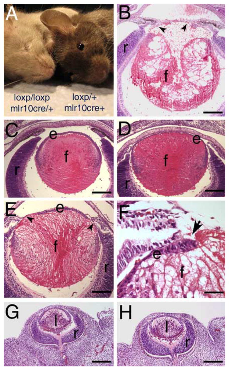
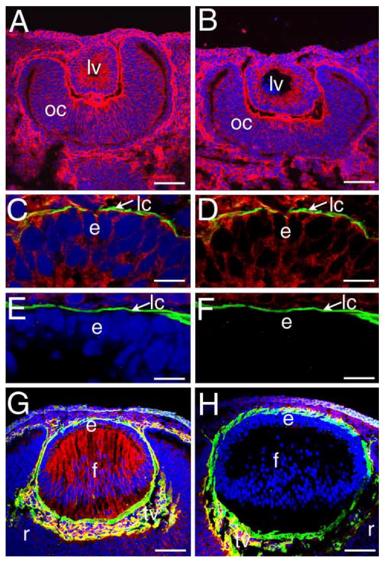
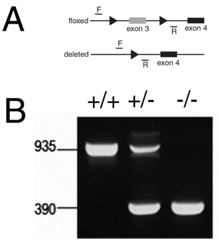
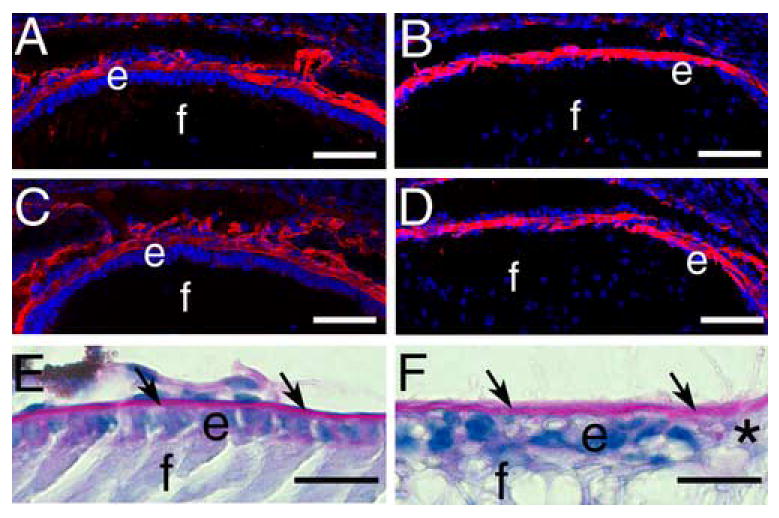
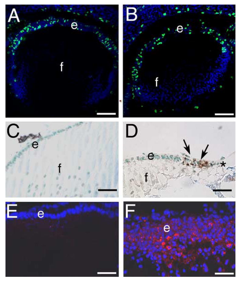
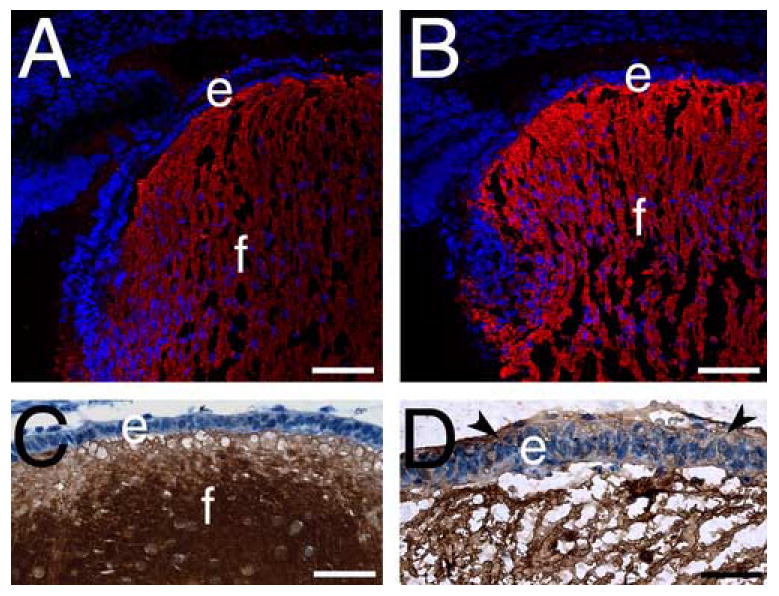
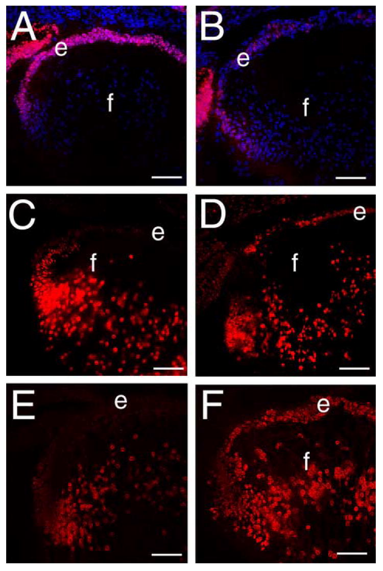
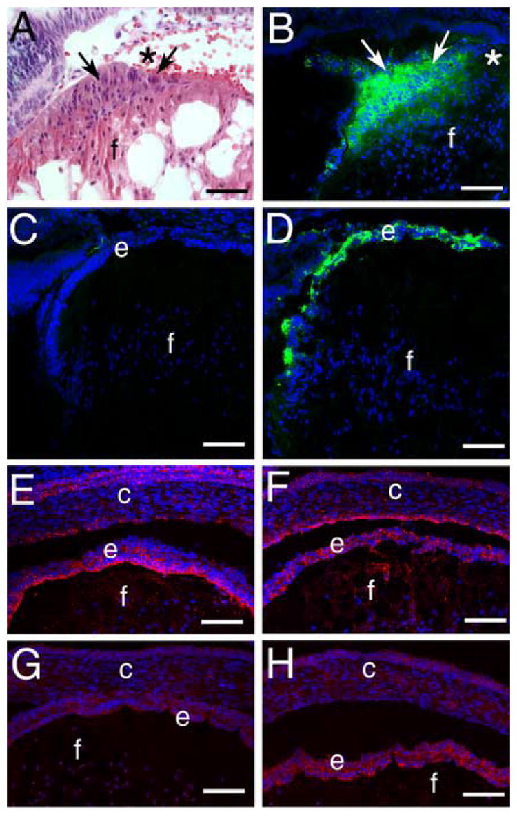
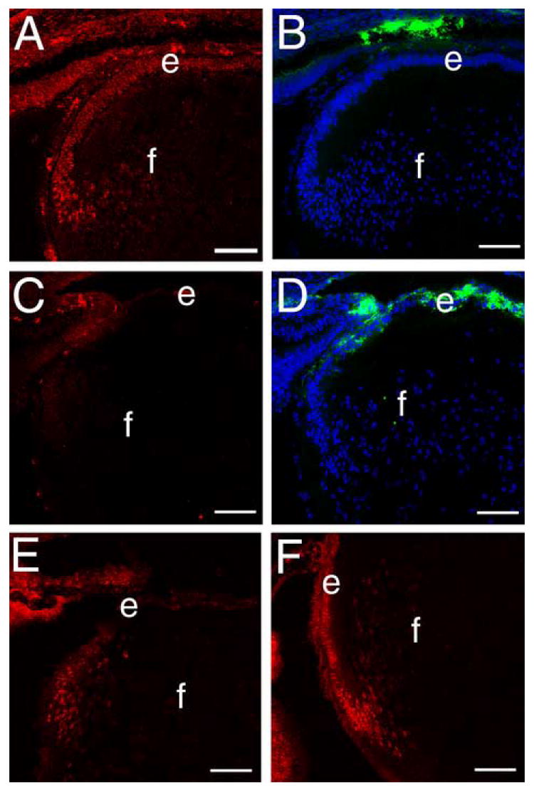
Beta1-integrins are cell surface receptors that participate in sensing the cell's external environment. We used the Cre-lox system to delete beta1-integrin in all lens cells as the lens vesicle transitions into the lens. Adult mice lacking beta1-integrin in the lens are microphthalmic due to apoptosis of the lens epithelium and neonatal disintegration of the lens fibers. The first morphological alterations in beta1-integrin null lenses are seen at 16.5 dpc when the epithelium becomes disorganized and begins to upregulate the fiber cell markers beta- and gamma-crystallins, the transcription factors cMaf and Prox1 and downregulate Pax6 levels demonstrating that beta1-integrin is essential to maintain the lens epithelial phenotype. Furthermore, beta1-integrin null lens epithelial cells upregulate the expression of alpha-smooth muscle actin and nuclear Smad4 and downregulate Smad6 suggesting that beta1-integrin may brake TGFbeta family signaling leading to epithelial-mesenchymal transitions in the lens. In contrast, beta1-integrin null lens epithelial cells show increased E-cadherin immunoreactivity which supports the proposed role of beta1-integrins in mediating complete EMT in response to TGFbeta family members. Thus, beta1-integrin is required to maintain the lens epithelial phenotype and block inappropriate activation of some aspects of the lens fiber cell differentiation program.


Figures

Figure 1
Figure 1
Mice homozygous for a floxed β1-integrin allele carrying MLR10-cre are microophthalmic with lens defects first apparent in late embryonic development. A) Exterior appearance of littermates who are either homozygous or heterozygous for floxed β1-integrin and carry MLR10-cre. B-H) Hematoxylin and eosin stained paraffin sections B) Newborn eye from a mouse homozygous for the floxed β1-integrin allele and carrying MLR10-cre showing loss of the anterior epithelium (arrowheads) and vacuolation of the lens fiber cells. C) Lens from a 16.5 dpc mouse homozygous for the floxed β1-integrin allele but lacking MLR10-CRE showing the normal regular organization of the lens epithelium and fiber cells. D) Eye from a 16.5 dpc mouse homozygous for the floxed β1-integrin allele and carrying MLR10-cre showing mild abnormalities in organization of the lens epithelium and fiber cells. E) Eye from 16.5 dpc mouse homozygous for the floxed β1-integrin allele and carrying MLR10-cre showing further elongation of the lens epithelial cells and regions of lens epithelial cell loss. F) High magnification image of a region of epithelial cell loss (arrow) observed in a 16.5 dpc lens. .Lens from 12.5 dpc mice either heterozygous (G) or homozygous (H) showing no abnormalities in lens morphology. Abbreviations; e, lens epithelium; f, lens fiber cells; r, retina; l- lens. Scale bars: Panels B, C, D, E, G, H-160 μm, Panel F- 40 μm
Figure 2
Figure 2
Homozygous flox β1-integrin/MLR10-cre mice lose β1 integrin protein from the lens by 11.5 dpc. A, B) Eyes from 10.5 dpc mice either heterozygous (A) or homozygous (B) for the floxed β1-integrin allele carrying MLR10-cre showing no changes in β1-integrin protein distribution (red) at the lens vesicle stage. C) The anterior lens vesicle of a 11.5 dpc wildtype mouse embryo triple labeled for β1-integrin (red), collagen IV (green) and DNA (blue). Panel D shows the same image as panel C with the DNA channel deleted. E) Anterior lens vesicle from a 11.5 dpc mouse embryo carrying the floxed β1-integrin allele and MLR10-cre triple labeled for β1-integrin (red), collagen IV (green) and DNA (blue). Panel F shows the same image as E with the DNA channel deleted. G) Eye from a wildtype 12.5 dpc mouse embryo showing the normal distribution of β1-integrin (red) in the lens at this stage. H) Eye from a 12.5 dpc mouse embryo homozygous for the floxed β1-integrin allele and carrying MLR10-cre showing the specific loss of β1-integrin protein from all lens cells. Red- β1-integrin, blue- DNA, green collagen IV; Panels A, B, G, H- Bar=77 μm; Panels C-F, Bar= 13 μm Abbreviations: lv- lens vesicle; oc- optic cup; e- lens epithelium; f- lens fibers; r- retina; lc-lens capsule; tv- tunica vasculosa.
Figure 3
Figure 3
Deletion of exon 3 from the β1-integrin gene using MLR10 CRE is efficient in the lens A) Diagram of the β1-integrin locus showing the postion of the PCR primers used to score the deletion and the presence of the loxP sites (arrowheads). B) PCR analysis of DNA obtained from newborn lenses either wildtype (+/+), heterozygous (+/-) or homozygous (-/-) for the floxed β1-integrin allele demonstrating that β1-integrin deletion is essentially complete.
Figure 4
Figure 4
β1-integrin null lenses retain the lens capsule A, B) Immunofluorescent staining of collagen IV (red) in the wildtype (A) and β1-integrin null (B) 16.5 dpc lens capsule C, D) Immunofluorescent staining of laminin (red) in the wildtype (C) and β1-integrin null (D) 16.5 dpc lens capsule. E, F) Periodic acid-Schiff staining (pink) of the newborn lens capsule (arrows) from wildtype (E) and β1-integrin null (F)mouse lenses showing that the lens capsule is retained even in areas denuded of lens epithelial cells (*). Abbreviations; e-lens epithelium; f- lens fiber cells. Panels A-D: scale bar= 77 μm. Panels E, F: scale bar =20 μm.
Figure 5
Figure 5
β1-integrin null lens epithelial cells are lost by birth due to elevated levels of apoptosis A, B) 16.5 dpc embryonic lenses stained for cells actively synthesizing DNA (green) using the 5-bromodeoxyuridine assay. Note that wildtype (A) and β1-integrin null (B) lens epithelia have qualitatively similar levels of cell proliferation. C, D) newborn lenses stained for apoptotic cells (brown) using TUNEL assay. Note that wildtype lenses (C) do not exhibit TUNEL positive lens epithelial cells while numerous TUNEL positive cells (arrows) are found in β1-integrin null lenses (D), especially in regions of lens epithelial cell loss (*). E, F) Newborn lens epithelial cells stained for cleaved caspase3 (Asp175). Note that wildtype lenses have little to no detectable cleaved caspase 3 (E) while numerous β1-integrin null lens epithelial cells are cleaved caspase 3 positive. Abbreviations: e- lens epithelium; f- lens fibers. Panels A,B: scale bar= 77 μm, C,D=30 μm, E,F=38 μm.
Figure 6
Figure 6
β-crystallin but not aquaporin0 expression is upregulated in 16.5 dpc β1-integrin null lens epithelial cells A,B) Aquaporin 0 (red) immunolocalization in wildtype (A) and β1-integrin (B) null lenses. C, D) β-crystallin (brown) immunolocalization in wildtype (C) and β1-integrin (D) null lenses. Abbreviations. e- lens epithelium, f- lens fibers. Scale Bar: Panels A,B= 77 μm; Panels C,D= 30 μm
Figure 7
Figure 7
β1-integrin null lens epithelial cells show downregulated Pax6 and upregulated cMaf and Prox1 expression at 16.5 dpc A, B Pax6 (red) immunolocalization in wildtype (A) and β1-integrin (B) null lenses, C,D) cMaf (red) immunolocalization in wildtype (C) and β1-integrin (D) null lenses. E, F) Prox1 (red) immunolocalization in wildtype (E) and β1-integrin (F) null lenses. Abbreviations: e-lens epithelium; f- lens fibers. Blue in panel A,B is DNA staining All scale bars= 77 μm
Figure 8
Figure 8
β1-integrin null lens epithelial cells demonstrate some but not all features of epithelial-mesenchymal transition (EMT). A) Hematoxylin and eosin stained paraffin section of the β1-integrin null newborn lens epithelium showing the elongation of these cells (arrows) from their normal flattened cuboidal morphology and the edge of the region of central epithelial cell loss (*). B) Immunodetection of αSMA (green) in the β1-integrin null newborn lens epithelium showing strong staining in the abnormal lens epithelial cells. C) Immunolocalization of αSMA (green) in the 16.5 dpc wildtype lens . D) Immunolocalization of αSMA (green) in the 16.5 dpc β1-integrin null lens imaged under the same conditions as panel C showing αSMA upregulation in the disorganized lens epithelium. E) Immunodetection of connexin 43 (red) in the wildtype 16.5 dpc lens demonstrating its preferred localization at the apical side of the lateral membranes of the lens epithelium. F) Immunodetection of connexin 43 (red) in the β1-integrin null 16.5 dpc lens demonstrating that its distribution is disorganized in the lens epithelium. G) Immunodetection of E-cadherin (red) in the wildtype 16.5 dpc lens demonstrating its expression in the lens epithelium. H) Immunodetection of E-cadherin (red) in the β1-integrin null 16.5 dpc lens demonstrating increased E-cadherin staining in the β1-integrin null lens epithelium. Abbreviations: c-cornea; e-lens epithelium; f-lens fiber cells scale bar: Panel A=30 μm, Panels B-H=77 μm
Figure 9
Figure 9
Smad6 expression is downregulated and Smad4 levels are upregulated in β1-integrin null lens epithelial cells A) Distribution of Smad6 (red) in the wildtype 16.5 dpc lens. B) the same section shown in A stained for DNA (blue) and αSMA (green). C) Distribution of Smad6 (red) in the β1-integrin null 16.5 dpc lens showing the downregulation of Smad6 levels in the lens epithelium D) the same section shown in C stained for DNA (blue) and αSMA (green) showing the upregulation of αSMA levels in a lens with reduced Smad6 levels. E) the distribution of Smad4 (red) in the wildtype 16.5 dpc lens demonstrating that this protein is predominately located in the nuclei of lens cells undergoing the transition between epithelial and fiber cells. F) The distribution of Smad4 (red) in the β1-integrin null 16.5 dpc lens showing that nuclear Smad4 levels are levated in β1-integrin null lens epithelial cells. Abbreviations: e-lens epithelium; f- lens fibers. All scale bars =77 μm

Similar articles

Cited by

References

    1. Ashery-Padan R, Marquardt T, Zhou X, Gruss P. Pax6 activity in the lens primordium is required for lens formation and for correct placement of a single retina in the eye. Genes Dev. 2000;14:2701–11. - PMC - PubMed
    1. Barbour W, Saika S, Miyamoto T, Ohkawa K, Utsunomiya H, Ohnishi Y. Expression patterns of beta1-related alpha integrin subunits in murine lens during embryonic development and wound healing. Curr Eye Res. 2004;29:1–10. - PubMed
    1. Bassnett S, Kuszak JR, Reinisch L, Brown HG, Beebe DC. Intercellular communication between epithelial and fiber cells of the eye lens. J Cell Sci. 1994;107(Pt 4):799–811. - PubMed
    1. Bassnett S, Missey H, Vucemilo I. Molecular architecture of the lens fiber cell basal membrane complex. J Cell Sci. 1999;112:2155–2165. - PubMed
    1. Belkin AM, Stepp MA. Integrins as receptors for laminins. Microsc Res Tech. 2000;51:280–301. - PubMed

Publication types

LinkOut - more resources