. 2007 Jul 1;313(11):2504-20.
doi: 10.1016/j.yexcr.2007.03.036. Epub 2007 Apr 6.

Prostaglandins PGE(2) and PGI(2) promote endothelial barrier enhancement via PKA- and Epac1/Rap1-dependent Rac activation

Affiliations

Prostaglandins PGE(2) and PGI(2) promote endothelial barrier enhancement via PKA- and Epac1/Rap1-dependent Rac activation

Anna A Birukova et al. Exp Cell Res. .

Abstract

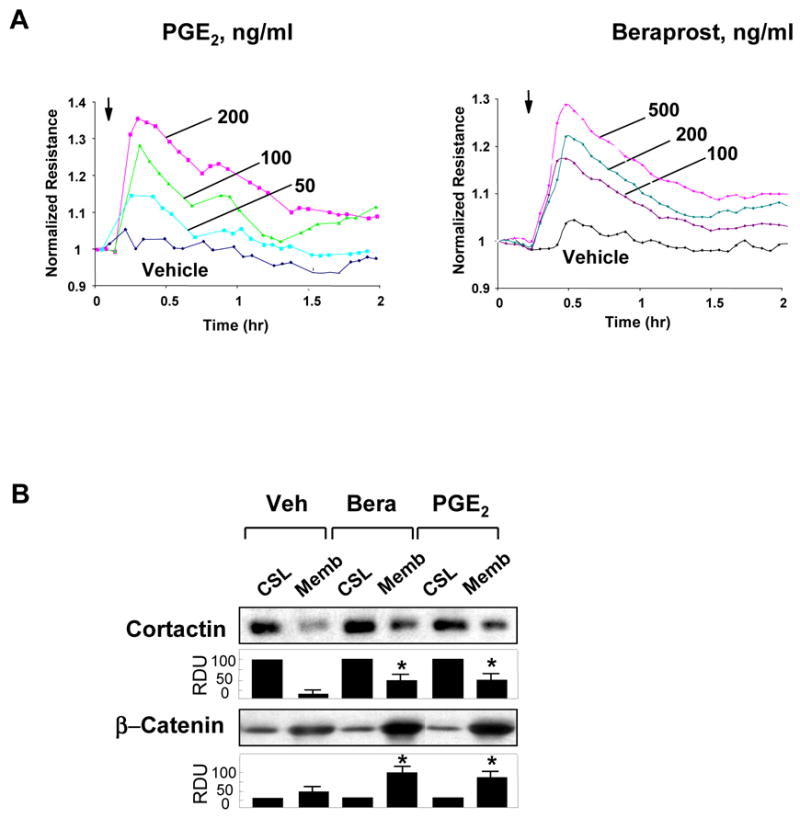
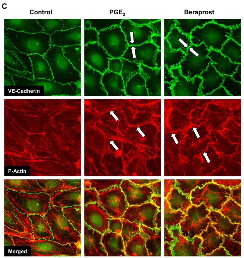
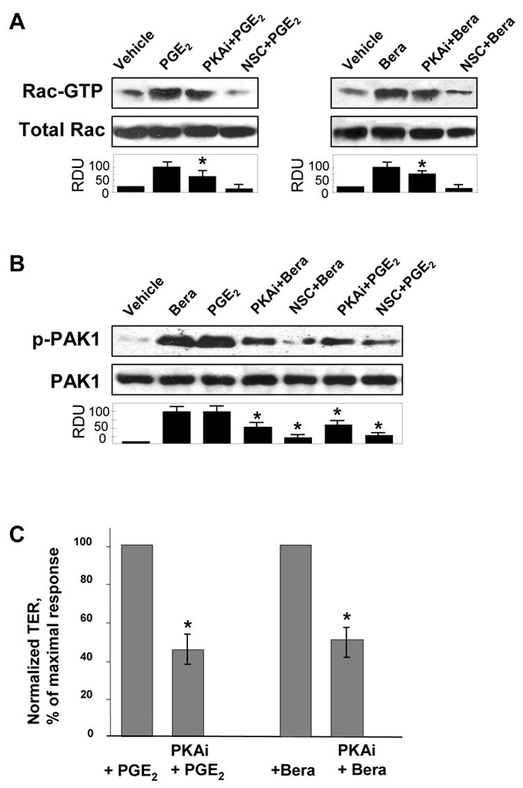
Prostaglandin E(2) (PGE(2)) and prostacyclin are lipid mediators produced by cyclooxygenase and implicated in the regulation of vascular function, wound repair, inflammatory processes, and acute lung injury. Although protective effects of these prostaglandins (PGs) are associated with stimulation of intracellular cAMP production, the crosstalk between cAMP-activated signal pathways in the regulation of endothelial cell (EC) permeability is not well understood. We studied involvement of cAMP-dependent kinase (PKA), cAMP-Epac-Rap1 pathway, and small GTPase Rac in the PGs-induced EC barrier protective effects and cytoskeletal remodeling. PGE(2) and PGI(2) synthetic analog beraprost increased transendothelial electrical resistance and decreased dextran permeability, enhanced peripheral F-actin rim and increased intercellular adherens junction areas reflecting EC barrier-protective response. Furthermore, beraprost dramatically attenuated thrombin-induced Rho activation, MLC phosphorylation and EC barrier dysfunction. In vivo, beraprost attenuated lung barrier dysfunction induced by high tidal volume mechanical ventilation. Both PGs caused cAMP-mediated activation of PKA-, Epac/Rap1- and Tiam1/Vav2-dependent pathways of Rac1 activation and EC barrier regulation. Knockdown of Epac, Rap1, Rac-specific exchange factors Tiam1 and Vav2 using siRNA approach, or inhibition of PKA activity decreased Rac1 activation and PG-induced EC barrier enhancement. Thus, our results show that barrier-protective effects of PGE(2) and prostacyclin on pulmonary EC are mediated by PKA and Epac/Rap pathways, which converge on Rac activation and lead to enhancement of peripheral actin cytoskeleton and adherens junctions. These mechanisms may mediate protective effects of PGs against agonist-induced lung vascular barrier dysfunction in vitro and against mechanical stress-induced lung injury in vivo.


Figures

Figure 1
Figure 1. Effect of PGE2 and beraprost on EC permeability, intracellular redistribution of cortactin and β-catenin and EC cytoskeletal remodeling
A: Human pulmonary EC monolayers were grown on gold microelectrodes. At the time point indicated by arrow, cells were treated with 50 ng/ml, 100 ng/ml, or 200 ng/ml PGE2 (left panel) or 100 ng/ml, 200 ng/ml, or 500 ng/ml beraprost (right panel) followed by measurements of TER reflecting EC monolayer barrier properties. B: EC were stimulated with PGE2 (200 ng/ml) or beraprost (500 ng/ml) for 15 min, and translocation of cortactin and β-catenin to the membrane fraction was detected with specific antibodies. Results are representative of three independent experiments. C: EC monolayers grown on glass coverslips were treated with PGE2 (200 ng/ml, 15 min) or beraprost (500 ng/ml, 15 min) followed by double immunofluorescent staining for VE-cadherin (upper panels) and F-actin (middle panels). Lower panel shows merged images of F-actin and VE-cadherin staining.
Figure 2
Figure 2. Effect of PGE2 or beraprost on activation of Rac-dependent signaling
EC were stimulated with PGE2 (200 ng/ml) or beraprost (500 ng/ml) for indicated periods of time. A: GTPase activation assays. Effects of PGE2 and beraprost on activation of Rac (upper panels), Cdc42 and Rho (lower panels) were evaluated using GTPase pulldown assays and normalized to the total GTPase content in cell lysates. B: Phosphorylation of Vav2 in control and PG-stimulated EC was determined in the total lysates using phospho-Vav2 specific antibody (left panels), or by immunofluorescent staining of beraprost-stimulated EC with phospho-Vav2 antibody (right panels). C: Time-dependent translocation of Tiam1 and p115-RhoGEF to the membrane fraction was detected by western blot with corresponding antibodies. D: Phosphorylation of PAK1 was determined in the total lysates using phospho-PAK1 specific antibody. Results are representative of three independent experiments.
Figure 3
Figure 3. Effect of PGE2 and beraprost on cAMP, cGMP, and PKA activation
A: EC were stimulated with PGE2 (200 ng/ml) or beraprost (500 ng/ml) for indicated periods of time, and intracellular cAMP and cGMP levels were determined using a non-radioactive immunoassay, as described in Materials and Methods. Results are mean ± SD of three independent experiments. *P<0.001. B and C: Cell lysates were analyzed for PKA activity by non-radioactive in vitro PKA assay. EC were stimulated with PGE2 (200 ng/ml) or beraprost (500 ng/ml) for indicated periods of time followed by determination of PKA activity (B). HPAEC were pretreated with GGT1 (30 μM, 1 hr) or PKI (20 μM, 1hr) prior to PGE2 or beraprost challenge for 5 min (C). The insets represent fluorescent phosphorylated form of PKA substrate kemptide separated from non-phosphorylated form by 0.8% agarose gel electrophoresis. The fluorescence intensity was detected and quantified by EagleEye Image System. Results are mean ± SD of three independent experiments. *P<0.001.
Figure 4
Figure 4. Effect of PKA inhibition on PGE2- and beraprost-induced Rac activation and EC barrier enhancement
A: Pulmonary EC were pretreated with PKA inhibitory peptide PKI (20 μM, 1hr) or Rac inhibitor NSC-23766 (200 μM, 1hr) prior to PGE2 (200 ng/ml) or beraprost (500 ng/ml) stimulation. Measurements of Rac activity were performed using pull-down assays. B: PAK1 phosphorylation was detected by immunoblotting with phospho-specific antibodies. C: Pulmonary EC were pretreated with PKI (20 μM, 1hr) prior to PGE2 (200 ng/ml) or beraprost (500 ng/ml) stimulation, and TER changes after 15 min corresponding to maximal EC response were measured. Results are representative of three to five independent experiments.
Figure 5
Figure 5. Effect of PGE2 and beraprost on Epac-Rap1 and Tiam1/Vav2-dependent Rac activation
A: Rap1 activation pull-down assay. EC were stimulated with PGE2 (200 ng/ml) or beraprost (500 ng/ml) for indicated periods of time. Upper panels depict Epac1 bound to activated Rap1 (Rap-GTP). Lower panel shows total Rap1 content in EC lysates. B: EC were preincubated with inhibitor of Rap1 processing GGT1 (30 μM, 1 hr) or vehicle followed by the measurements of PGE2-induced Rac activation. C: Pulmonary EC were transfected with siRNA specific to Epac1, Rap1, Tiam1, or Vav2. Depletion of target proteins induced by specific siRNA duplexes was confirmed by immunoblotting with appropriate antibody, as compared to treatment with non-specific RNA. Immunoblot with β-actin antibody was used as normalization control. Results are representative of three to five independent experiments. D: Pulmonary EC were transfected with specific siRNAs followed by agonist stimulation and measurements of Rac activity. Control cells were treated with non-specific RNA. Results are representative of three to six independent experiments.
Figure 6
Figure 6. Involvement of Rac, Epac1, Rap1, Tiam1, Vav2, and PKA in prostaglandin-mediated EC barrier enhancement
A: EC monolayers were transfected with siRNA specific to Rap1, Tiam1, Vav2, Rac, or GEF-H1. Control cells were transfected with non-specific RNA. After 72 hrs of transfection, cells were stimulated with PGE2 (200 ng/ml), and TER changes after 15 min corresponding to maximal EC response were measured. B: Cells were transfected with Epac1-specific or non-specific siRNA. Measurements of EC permeability were performed in EC stimulated with PGE2 (200 ng/ml) or beraprost (500 ng/ml), or pre-treated with PKI (20 μM, 1hr) prior to PGs stimulation. Bar graphs depict changes in TER determined after 15 min of PG stimulation. Results are representative of three to seven independent experiments.
Figure 7
Figure 7. Effect of Rap1 and Rac knockdown on PGE2-induced cytoskeletal and adherens junction remodeling
EC grown on glass coverslips and transfected with non-specific RNA (A), with Rap1-specific siRNA (B) or with Rac-specific siRNA (C) were stimulated with PGE2 (200 ng/ml, 15 min). Analysis of actin cytoskeletal remodeling was performed by double immunofluorescent staining with VE-cadherin and Texas Red phalloidin, as described in Methods section. Lower panel shows merged images of F-actin and VE-cadherin staining.
Figure 8
Figure 8. Protective effects of prostaglandins in pulmonary cell culture model of thrombin-induced hyper-permeability and murine model of VILI
A: Effect of prostaglandins on thrombin-induced barrier dysfunction. Pulmonary EC were pre-incubated with PGE2 (200 ng/ml) or beraprost (500 ng/ml) for 15 min followed by thrombin (0.5 U/ml) challenge, and TER changes were monitored over the time. B: Effect of prostaglandins on thrombin-induced EC permeability. Pulmonary EC were pre-incubated with PGE2 (200 ng/ml) or beraprost (500 ng/ml) followed by thrombin (0.5 U/ml) challenge, and measurements of permeability for FITC-labeled dextran. C: Effect of PG (beraprost, 500 ng/ml, 15 min) on thrombin (0.5 U/ml, 15 min)-induced cytoskeletal remodeling and adherens junction integrity. Double immunofluorescence staining was performed using VE-cadherin antibodies and Texas-Red phalloidin. Paracellular gaps are marked by arrows. D and E: Effect of PG on thrombin-induced activation of Rho-dependent pathway. HPAEC were pre-incubated with beraprost followed by treatment with thrombin for 5 min or 30 min and determination of Rho activity using pull-down assay (D). Phosphorylation of MLC in EC pretreated with beraprost followed by thrombin challenge was detected by western blot with specific antibodies (E). F: − 57BL/6J mice were subjected to mechanical ventilation at high tidal volume (HTV, 30 ml/kg, 4 hrs) or left spontaneously ventilated. Intravenous administration of beraprost (2 μg/kg) was performed at three time points (0, 40, and 80 min) during mechanical ventilation. Cell count (upper panel) and measurement of protein concentration (lower panel) were performed in bronchoalveolar lavage fluid taken from control and experimental animals. Results are represented as mean + SE; *p < 0.01; **p < 0.05; n=6 per group.
Figure 9
Figure 9. Upstream mechanisms of PG-induced Rac activation and cytoskeletal remodeling leading to endothelial barrier enhancement
Stimulation of EC with PGE2 or PGI2 elevates intracellular cAMP levels and stimulates cAMP-dependent protein kinase (PKA), cAMP-activated guanine nucleotide exchange factor Epac1, which activates its effector small GTPase Rap1. Activated PKA and Rap1 promote Rac activation via stimulation of Rac specific GEFs Tiam1 and Vav2. Activated Rac interacts with downstream cytoskeletal and cell adhesion effectors and promotes cytoskeletal remodeling and EC barrier enhancement. In addition, PKA may directly affect EC cytoskeletal organization and monolayer barrier properties via modulation of myosin light chain kinase activity or VASP-dependent relaxation of actin cytoskeleton.

References

    1. Ricard JD, Dreyfuss D, Saumon G. Ventilator-induced lung injury. Eur Respir J Suppl. 2003;42:2s–9s. - PubMed
    1. Matthay MA, Zimmerman GA, Esmon C, et al. Future research directions in acute lung injury: summary of a National Heart, Lung, and Blood Institute working group. Am J Respir Crit Care Med. 2003;167:1027–35. - PubMed
    1. Park GY, Christman JW. Involvement of cyclooxygenase-2 and prostaglandins in the molecular pathogenesis of inflammatory lung diseases. Am J Physiol Lung Cell Mol Physiol. 2006;290:L797–805. - PMC - PubMed
    1. Howard LS, Morrell NW. New therapeutic agents for pulmonary vascular disease. Paediatr Respir Rev. 2005;6:285–91. - PubMed
    1. Ueno Y, Koike H, Annoh S, et al. Anti-inflammatory effects of beraprost sodium, a stable analogue of PGI2, and its mechanisms. Prostaglandins. 1997;53:279–89. - PubMed

Publication types

MeSH terms