. 2007 Jun;52(8):1631-40.
doi: 10.1016/j.neuropharm.2007.03.010. Epub 2007 Mar 24.

Presynaptic AMPA and kainate receptors increase the size of GABAergic terminals and enhance GABA release

Affiliations

Presynaptic AMPA and kainate receptors increase the size of GABAergic terminals and enhance GABA release

Mónica L Fiszman et al. Neuropharmacology. 2007 Jun.

Abstract

In the developing cerebellum, NMDA receptors promote the maturation of axonal terminals of inhibitory interneurons. We compared the effects of AMPA/kainate receptor agonists in cultured cerebellar cells from GAD65-eGFP mice. Both AMPA and kainate augmented granule cell survival without affecting interneurons. The action of kainate was blocked by an AMPA but not by a NMDA receptor antagonist, suggesting AMPA receptor involvement. AMPA and kainate increased the size of the GABAergic terminals and the action of kainate was insensitive to NMDA blockers. Whole cell recordings in granule neurons revealed that chronic treatment for 5 days with kainate as well as NMDA decreased AMPA receptor expression while interneuronal kainate receptors were depressed by kainate treatment. Acute kainate application increased mIPSCs frequency in both granule neurons and interneurons and this effect was only partially blocked by an AMPA receptor antagonist. In contrast to what was reported for NMDA, chronic treatment with kainate induced a significant decrease of the basal mIPSCs frequency but increased the acute action of kainate on mIPSCs. Direct recordings from presynaptic GABAergic terminals suggest that AMPA and kainate receptors are present in developing GABAergic terminals and their activation affects the size of GABAergic terminals and spontaneous GABA release.


Figures
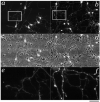
Fig.1
Fig.1. Cerebellar neurons in primary cultures from GAD-65 expressing mice
Cerebellar cultures at DIV7 grown for 5 days in the absence (left) or the presence of 20 μM kainate (right). Qualitative observations of fluorescent images indicate substantial changes induced by kainate treatment: increased survival of granule neurons and enhanced size of the GABAergic axonal varicosities. In panels c and d the fluorescence microphotographs in a and b are overlapped to phase contrast ones to illustrate the density of granule neurons and GABAergic interneurons. Panels e and f illustrate a magnified sections of panels a and b that better show GABAergic axonal branches and presynaptic varicosities. Calibration bar 30 μm and 6 μm for the insets.
Fig. 2
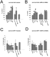
Fig. 2. AMPA & NMDA receptors increase survival of granule neurons in cerebellar cultures
A,B Survival of cerebellar neurons was increased by treatments for 5 days with increasing doses of kainate (K, A) and AMPA (A, B). Combination of these treatments with the selective AMPA receptor antagonist GYKI52466 (GYKI, 25 μM) completely blocked this action. In contrast, blockade with the NMDA receptor antagonist MK801 (MK, 10 μM) failed to reverse kainate action. In B is also shown that GYKI52466 (25 μM) failed to reverse the reported facilitation of survival with NMDA (N, 100 μM) treatment. Cyclothiazide (Cyclo, 100 μM) failed to enhance the action of AMPA. C,D Survival of eGFP positive neurons expressed as % of total number of cells in each field analyzed. Although both treatments increased the total number of neurons and therefore the absolute number of eGFP labeled GABAergic interneurons, the percent of these neurons was not increased by treatments with increasing doses of both kainate (A) and AMPA (B) in contrast to treatments with NMDA. Data derive from at least 6 coverslips from four distinct cultures and are expressed as mean ± SD. * significant to control, p<0.01. + significant to K20+GYKI, p<0.01.
Fig.3
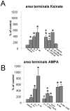
Fig.3. Kainate and AMPA enlarge GABAergic terminals
Summary results on the analysis of eGFP labeled varicosities in axons of cerebellar GABAergic interneurons grown in distinct culture conditions. A: The percentage increase in the area of the varicosities induced by 0.2, 2, and 20 μM Kainate is compared to the increase obtained in the presence of kainate (K, 20 μM) together with GYKI52466 (GYKI, 25 μM or 50 μM) or MK801 (MK, 10 μM). No change in terminal size was seen with GYKI52466 (GYKI, 50 μM) alone. The area of varicosities in the vehicle group was 0.75 ± 0.18 μm2. * significant to vehicle, p<0.01. + significant to K20, p<0.01. B: The steep increase in the area of varicosities caused by increasing doses of AMPA (A, 0.2, 2 and 10 μM) is compared to that of NMDA (N, 100 μM). GYKI52466 blocked AMPA but not NMDA-induced increase in the size of the varicosities. In addition it is shown the effect on varicosity enlargement of cyclothiazide (Cyclo, 100 μM) a drug that selectively potentiates the action of flip variants of AMPA receptors. Data derive from at least 30 segments in each experimental group from 6 separate cultures and are expressed as mean ± SD. * significant to vehicle, p<0.01. + significant to A 0.2, p<0.01. × significant to A 2, p<0.01.
Fig. 4
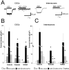
Fig. 4. Whole cell current elicited by kainate in CGCs and interneurons
Effect of direct application of kainate to cerebellar granule neurons and interneurons at DIV 7-8. A: Comparison of representative whole cell current records from a granule cell (left) and an interneuron (right) at DIV7, voltage clamped at -60 mV. Horizontal bars correspond to the acute application of kainate (20 μM, K20) alone or in combination with GYKI (50 μM, K20+GYKI). The average whole cell current density obtained with applications of 20 μM kainate in cerebellar granule neurons (B) or interneurons (C) is reported for cells in control conditions or cells treated for 5 days with kainate (20 μM) or NMDA (100 μM). Also included is a comparison of whole cell current density obtained with 2 μM kainate (K2), 2 μM kainate + 20 μM Con A (K2+conA) and 20 μM kainate + 50 μM GYKI 52466 (K20+GYKI) in cerebellar granule neurons (B) or interneurons (C) in control conditions or treated for 5 days with kainate (20 μM) or NMDA (100 μM). Data derive from at least 15 granule neurons and 16 interneurons in 5 distinct sets of cultures at DIV7-8 for vehicle and kainate treatment and from 6 granule neurons and 5 interneurons for NMDA treatments. * significant to vehicle, p<0.01. + significant to K20, p<0.01 × significant to K2 application, p<0.01.
Fig. 5
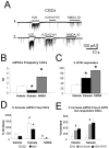
Fig. 5. increase of mIPSCs by kainate is altered by chronic treatments with kainate and NMDA
Effect of chronic and acute application of kainate and NMDA on GABAergic mIPSCs recorded from cerebellar granule cells at DIV 7-8. A. Representative miniature inhibitory postsynaptic currents (mIPSCs) records from cerebellar granule neurons at DIV 7 grown in vehicle, control conditions. Horizontal bar corresponds to the acute application of kainate (K, 20 μM K20) alone or together with GYKI52466 (50 μM, K20+GYKI). The effect of NMDA (NMDA, 30 μM) is also illustrated. Recordings from cells 1 & 2 differ dramatically in the action of GYKI52466 suggesting the presence of presynaptic kainate receptors in cell 2. B. A comparison of the average mIPSCs frequency in granule neurons at DIV7 grown in vehicle or chronically treated with kainate (20 μM) or NMDA (100 μM) for 5 days. * p<0.01 compared to vehicle. C. A comparison of the percent of cells that did not display increase in mIPSC frequency with coapplication of 20 μM kainate and 50 μM GYIK52466 (%GYKI) in granule neurons at DIV7 grown in control conditions or chronically treated with kainate (20 μM) or NMDA (100 μM) for 5 days. Data derive from 15 granule neurons in 5 different cultures and averaged across distinct cultures * p<0.01 compared to vehicle. D. Comparison of the % increase of mIPSC frequency by acute applications of 20 μM kainate (K20) with NMDA (N, 30 μM) (Left panel) in control cells as compared to cells chronically treated with kainate (20 μM) or NMDA (100 μM) for 5 days. * significant to vehicle treated cells, p<0.01. Data from at least 15 granule neurons in 5 distinct sets of cultures at DIV7-8 for control and kainate treatment and from 6 granule neurons and 7 interneurons for NMDA treatments. E. Comparison of the % increase of mIPSC frequency by acute applications of 2 μM kainate with 20 μM kainate + 50 μM GYKI52466 (K20+GYKI,) or 2 μM kainate after pretreatment with ConA 20 μM (K2+conA) in cells that responded with mIPSCs frequency increase even when kainate and GYKI52466 were combined (GYKI non responder cells). Vehicle treated cultures were compared to cultures chronically treated with kainate (20 μM) for 5 days. × significant to K2 application, p<0.01. Data derives from at least 6 cells in two separate cultures
Fig. 6
Fig. 6. Kainate current recordings from presynaptic terminals
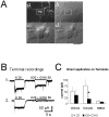
A. Microphotographs of cerebellar neurons at DIV8 in a kainate treated culture. Panel a shows GFP positive axons with large varicosities. The same field is shown to the right with DIC-Nomarski optics (panel b). White box inserts are magnified below showing a varicosity (GFP/DIC-Nomarski, panel c) in close proximity to the cell body of a granule neuron. The tip of a patch pipette touching the varicosity is shown in panel d. Calibration bar 30 μm (a,b) and 7 μm (c,d). B. Voltage-clamp current recording from two distinct GFP labeled varicosities in kainate treated cells illustrating kainate-elicited current and the variability of antagonism by the AMPA receptor selective antagonist GYKI52466. C. Summary of whole-terminal current activated by kainate (K) at 20 μM, or kainate 20 μM+ GYKI52466 (K20+GYKI, 50 μM) in control cells as compared to cells chronically treated with kainate (20 μM) or NMDA (100 μM) for 5 days. + p<0.05 compared to kainate (K) at 20 μM. Data derive from >10 terminals in 2 distinct sets of cultures at DIV7-8 for control and kainate treatment and from 5 terminals for NMDA treatments.

Similar articles

Cited by

References

    1. Baude A, Molnar E, Latawiec D, McIlhinney RA, Somogyi P. Synaptic and nonsynaptic localization of the GluR1 subunit of the AMPA-type excitatory amino acid receptor in the rat cerebellum. Journal of Neuroscience. 1994;14:2830–2843. - PMC - PubMed
    1. Bettler B, Egebjerg J, Sharma G, Pecht G, Hermans-Borgmeyer I, Moll C, Stevens CF, Heinemann S. Cloning of a putative glutamate receptor: a low affinity kainate-binding subunit. Neuron. 1992;8:257–265. - PubMed
    1. Brager DH, Luther PW, Erdelyi F, Szabo G, Alger BE. Regulation of exocytosis from single visualized GABAergic boutons in hippocampal slices. Journal of Neuroscience. 2003;23:10475–10486. - PMC - PubMed
    1. Bureau I, Mulle C. Potentiation of GABAergic synaptic transmission by AMPA receptors in mouse cerebellar stellate cells: changes during development. Journal of Physiology (London) 1998;509:817–831. - PMC - PubMed
    1. Damgaard I, Trenkner E, Sturman JA, Schousboe A. Effect of K+- and kainate-mediated depolarization on survival and functional maturation of GABAergic and glutamatergic neurons in cultures of dissociated mouse cerebellum. Neurochemical Research. 1996;21:267–275. - PubMed

Publication types

MeSH terms