. 2007 Sep 1;365(2):435-45.
doi: 10.1016/j.virol.2007.04.015. Epub 2007 May 9.

Cell cycle arrest and apoptosis induced by the coronavirus infectious bronchitis virus in the absence of p53

Affiliations

Cell cycle arrest and apoptosis induced by the coronavirus infectious bronchitis virus in the absence of p53

Frank Q Li et al. Virology. .

Abstract

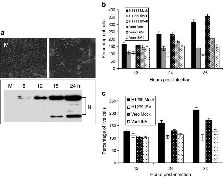
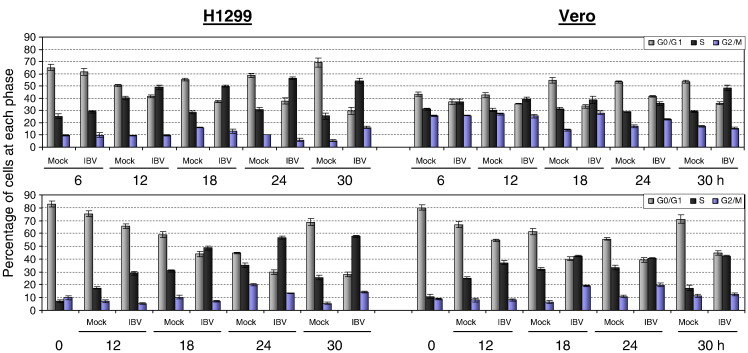
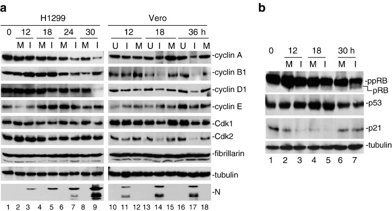
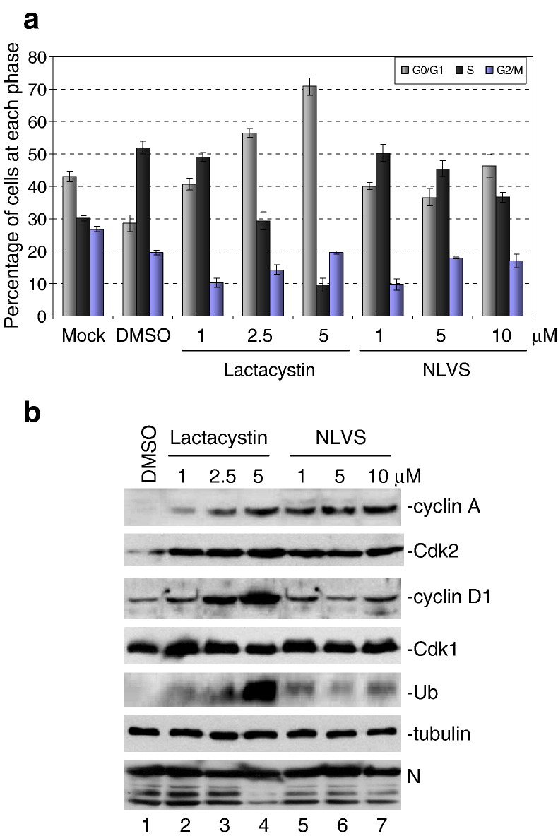
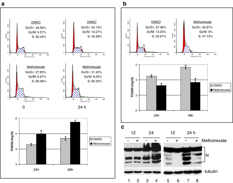
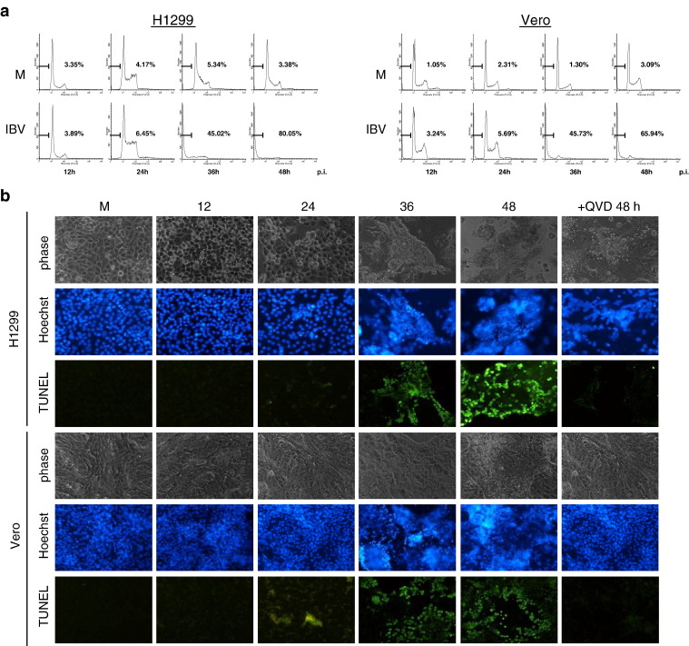
Manipulation of the cell cycle and induction of apoptosis are two common strategies used by many viruses to regulate their infection cycles. In cells infected with coronaviruses, cell cycle perturbation and apoptosis were observed in several reports. However, little is known about how these effects are brought out, and how manipulation of the functions of host cells would influence the replication cycle of coronavirus. In this study, we demonstrate that infection with coronavirus infectious bronchitis virus (IBV) imposed a growth-inhibitory effect on cultured cells by inducing cell cycle arrest at S and G(2)/M phases in both p53-null cell line H1299 and Vero cells. This cell cycle arrest was catalyzed by the modulation of various cell cycle regulatory genes and the accumulation of hypophosphorylated RB, but was independent of p53. Proteasome inhibitors, such as lactacystin and NLVS, could bypass the IBV-induced S-phase arrest by restoring the expression of corresponding cyclin/Cdk complexes. Our data also showed that cell cycle arrest at both S- and G(2)/M-phases was manipulated by IBV for the enhancement of viral replication. In addition, apoptosis induced by IBV at late stages of the infection cycle in cultured cells was shown to be p53-independent. This conclusion was drawn based on the observations that apoptosis occurred in both IBV-infected H1299 and Vero cells, and that IBV infection did not affect the expression of p53 in host cells.


Figures

Fig. 1
Fig. 1
Inhibition of cell proliferation in IBV-infected H1299 and Vero cells. (a) H1299 cells were mock (M) or infected with IBV (I) at an MOI of 1. The morphological characteristics of the infected cells were observed under a light microscope at 36 h post-infection (upper panels). At the indicated times, cells were lysed with SDS sample buffer and viral protein expression was tested by Western blot analysis with anti-IBV N polyclonal antibodies. (b) H1299 and Vero cells at 50% confluence in 6-well plates were infected with IBV at an MOI of 0.5 (IBV-I) or 1 (IBV-II). At various times post-infection, the total cell numbers were counted and plotted. Data are presented as percentage of cell numbers compared with that of mock-infected cells at 0 h post-infection. The percentages are results of five repeated experiments. (c) Analysis of cell proliferation by MTT assay. H1299 and Vero cells at 50% confluence in 96-well plates were mock- or IBV-infected at an MOI of 1. At various times post-infection, MTT assays were performed. Data are presented as percentage of live cells compared with that of mock-infected cells at 0 h post-infection. The percentages are results of five repeated experiments.
Fig. 2
Fig. 2
Induction of aberrant cell cycle progression in IBV-infected cells. H1299 and Vero cells were infected with IBV at an MOI of 1. At the indicated times, cells were collected and stained with propidium iodide for FACS analysis. Data were analyzed by using the ModFit LT Mac 3.0 software to determine the percentage of cells at each phase of the cell cycle in both asynchronously growing (upper panels) and synchronized (lower panels) H1299 and Vero cells. The results are presented as means of five repeated experiments.
Fig. 3
Fig. 3
Western blot analysis of cell cycle-related genes. (a) H1299 (left panels) and Vero (right panels) cells were either uninfected (U), mock-infected (M) or infected with IBV (I) at an MOI of 1. At the indicated times, cells were lysed with SDS sample buffer, and equal amounts of proteins from the samples were tested by Western blot analysis with anti-cyclins A, B1, D1, and E, Cdk1, Cdk2, and fibrillarin antibodies, respectively. The same membranes were also probed with β-tubulin as a loading control. Viral replication was confirmed by Western analysis of N protein with anti-IBV N polyclonal antibodies. The data were representatives of three repeated experiments. (b) Asynchronously growing H1299 (top panel) and Vero (other panels) cells were infected with IBV at an MOI of 1 (I). At the indicated times, cells were lysed and subjected to Western blot analysis with anti-RB, p53, p21 and β-tubulin antibodies, respectively. Hypophosphorylated forms of RB (pRB) appeared as fast-migrating bands, and hyperphosphorylated RB (ppRB) appeared as slightly retarded bands (top panel). The data are representative of three independent experiments.
Fig. 4
Fig. 4
Bypass of IBV-induced S-phase arrest in cells treated with proteasome inhibitors. (a) Vero cells were preincubated with either DMSO or increasing concentrations of proteasome inhibitors, lactacystin and NLVS, for 30 min and then infected with IBV. At 24 h post-infection, the cells were collected and subjected to FACS analysis. The data are presented as means of three repeated experiments. (b) Cell lysates were analyzed for cyclin A, D1, Cdk1, Cdk2, ubiquitin, β-tubulin, and IBV N protein by Western blotting. The data are representative of three independent experiments.
Fig. 5
Fig. 5
S and G2/M arrest promotes IBV replication. (a) Asynchronously growing H1299 cells were treated with DMSO or methotrexate for 20 h, followed by incubation in fresh medium for 6 h to release the cell cycle progression. Cells were then infected with IBV at an MOI of 1 and harvested at 24 and 48 h post-infection, and viral titers were determined (bottom panel). The cell cycle profiles at the end of fresh medium incubation and 24 h post-infection were analyzed by flow cytometric analysis (top and middle panels). (b) Asynchronously growing H1299 cells were mock-infected or infected with IBV at an MOI of 1, followed by incubation with either DMSO or methotrexate. The cell cycle profiles at 24 h post-infection were obtained by flow cytometric analysis (upper panels). Viral titers at 24 and 48 h post-infection were determined (lower panel). (c) Viral protein expression at 12 and 24 h post-infection was analyzed by Western blot with anti-IBV N polyclonal antibodies.
Fig. 6
Fig. 6
Induction of apoptotic cell death in IBV-infected H1299 and Vero cells. (a) H1299 and Vero cells were infected with IBV at an MOI of 1. At the indicated times post-infection, cells were collected and stained with propidium iodide for flow cytometric analysis. Cells with sub-G0/G1 (hypodiploid) DNA contents are indicated by a bar and as percentage in each graph. The data are representative of three independent experiments. (b) Morphological characteristics and TUNEL assays of mock- and IBV-infected H1299 and Vero cells at 12, 24, 36, and 48 h post-infection. Cells were mock- or IBV-infected, stained with Hoechst 33342 or analyzed by TUNEL assay with an in situ cell death detection kit at the indicated times post-infection, and viewed under a fluorescence microscope. At 48 h post-infection, IBV-infected cells treated with a caspase inhibitor, QVD, were also analyzed by the same assays. The data are representative of three independent experiments. Phase, phase-contrast images; Hoechst, nuclear staining; TUNEL, TUNEL assay.

Similar articles

Cited by

References

    1. An S., Chen C.-J., Yu X., Leibowitz J.L., Markino S. Induction of apoptosis in murine coronavirus-infected cultured cells and demonstration of E protein as an apoptosis inducer. J. Virol. 1999;73:7853–7859. - PMC - PubMed
    1. Barber G.N. Host defense, viruses and apoptosis. Cell Death Differ. 2001;8:113–126. - PubMed
    1. Benedict C.A., Norris P.S., Ware C.F. To kill or be killed: viral evasion of apoptosis. Nat. Immunol. 2002;3:1013–1018. - PubMed
    1. Chau T.N., Lee K.C., Yao H., Tsang T.Y., Chow T.C., Yeung Y.C., Choi K.W., Tso Y.K., Lau T., Lai S.T., Lai C.L. SARS-associated viral hepatitis caused by a novel coronavirus: report of three cases. Hepatology. 2004;39:302–310. - PMC - PubMed
    1. Chen C.-J., Makino S. Murine coronavirus replication induces cell cycle arrest in G0/G1 phase. J. Virol. 2004;78:5658–5669. - PMC - PubMed

Substances