Aggregation of TMV CP plays a role in CP functions and in coat-protein-mediated resistance
- PMID: 17493658
- PMCID: PMC2034504
- DOI: 10.1016/j.virol.2007.03.014
Aggregation of TMV CP plays a role in CP functions and in coat-protein-mediated resistance
Abstract
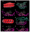
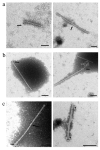
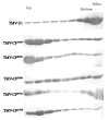
Tobacco mosaic virus (TMV) coat protein (CP) in absence of RNA self-assembles into several different structures depending on pH and ionic strength. Transgenic plants that produce self-assembling CP are resistant to TMV infection, a phenomenon referred to as coat-protein-mediated resistance (CP-MR). The mutant CP Thr42Trp (CP(T42W)) produces enhanced CP-MR compared to wild-type CP. To establish the relationship between the formation of 20S CP aggregates and CP-MR, virus-like particles (VLPs) produced by TMV variants that yield high levels of CP-MR were characterized. We demonstrate that non-helical structures are found in VLPs formed in vivo by CP(T42W) but not by wild-type CP and suggest that the mutation shifts the intracellular equilibrium of aggregates from low to higher proportions of non-helical 20S aggregates. A similar shift in equilibrium of aggregates was observed with CP(D77R), another mutant that confers high level of CP-MR. The mutant CP(D50R) confers a level of CP-MR similar to wild-type CP and aggregates in a manner similar to wild-type CP. We conclude that increased CP-MR is correlated with a shift in intracellular equilibrium of CP aggregates, including aggregates that interfere with virus replication.
Figures


Similar articles
-
Conformational behavior of coat protein in plants and association with coat protein-mediated resistance against TMV.Braz J Microbiol. 2020 Sep;51(3):893-908. doi: 10.1007/s42770-020-00225-0. Epub 2020 Jan 13. Braz J Microbiol. 2020. PMID: 31933177 Free PMC article.
-
Characterization of mutant tobacco mosaic virus coat protein that interferes with virus cell-to-cell movement.Proc Natl Acad Sci U S A. 2002 Mar 19;99(6):3645-50. doi: 10.1073/pnas.062041499. Epub 2002 Mar 12. Proc Natl Acad Sci U S A. 2002. PMID: 11891326 Free PMC article.
-
Studies of coat protein-mediated resistance to tobacco mosaic tobamovirus: correlation between assembly of mutant coat proteins and resistance.J Virol. 1997 Oct;71(10):7942-50. doi: 10.1128/JVI.71.10.7942-7950.1997. J Virol. 1997. PMID: 9311885 Free PMC article.
-
Coat-protein-mediated resistance to tobacco mosaic virus: discovery mechanisms and exploitation.Philos Trans R Soc Lond B Biol Sci. 1999 Mar 29;354(1383):659-64. doi: 10.1098/rstb.1999.0418. Philos Trans R Soc Lond B Biol Sci. 1999. PMID: 10212946 Free PMC article. Review.
-
Molecular assembly of tobacco mosaic virus in vitro.Adv Biophys. 1986;22:95-149. doi: 10.1016/0065-227x(86)90004-3. Adv Biophys. 1986. PMID: 3551520 Review.
Cited by
-
Stability of plasmid and viral banks supporting the cGMP manufacture of Q-Griffithsin from a TMV-based viral vector.J Biotechnol. 2020 Aug 20;320:74-76. doi: 10.1016/j.jbiotec.2020.06.009. Epub 2020 Jun 15. J Biotechnol. 2020. PMID: 32553533 Free PMC article.
-
Tomato plant cell death induced by inhibition of HSP90 is alleviated by Tomato yellow leaf curl virus infection.Mol Plant Pathol. 2016 Feb;17(2):247-60. doi: 10.1111/mpp.12275. Epub 2015 Jun 7. Mol Plant Pathol. 2016. PMID: 25962748 Free PMC article.
-
Strategies for antiviral resistance in transgenic plants.Mol Plant Pathol. 2008 Jan;9(1):73-83. doi: 10.1111/j.1364-3703.2007.00447.x. Mol Plant Pathol. 2008. PMID: 18705886 Free PMC article. Review.
-
Isolation of Tobacco Mosaic Virus-Binding Peptides for Biotechnology Applications.Chembiochem. 2022 Jun 3;23(11):e202200040. doi: 10.1002/cbic.202200040. Epub 2022 Apr 5. Chembiochem. 2022. PMID: 35320626 Free PMC article.
-
Agrobacterium rhizogenes-induced soybean hairy roots versus Soybean mosaic virus (ARISHR-SMV) is an efficient pathosystem for studying soybean-virus interactions.Plant Methods. 2019 May 25;15:56. doi: 10.1186/s13007-019-0442-8. eCollection 2019. Plant Methods. 2019. PMID: 31149022 Free PMC article.
References
-
- Asselin A, Zaitlin M. Characterization of a second protein associated with virions of tobacco mosaic virus. Virology. 1978;91(1):173–81. - PubMed
-
- Bazzini AA, Asurmendi S, Hopp HE, Beachy RN. Tobacco mosaic virus (TMV) and potato virus X (PVX) coat proteins confer heterologous interference to PVX and TMV infection, respectively. J Gen Virol. 2006;87(Pt 4):1005–12. - PubMed
-
- Beachy RN, Zaitlin M. Characterization and in vitro translation of the RNAs from less-than-full-length, virus-related, nucleoprotein rods present in tobacco mosaic virus preparations. Virology. 1977;81(1):160–9. - PubMed
Publication types
MeSH terms
Substances
Grants and funding
LinkOut - more resources
Full Text Sources
Miscellaneous

