Review
. 2007;120 Suppl 1(Suppl 1):S5-9.
doi: 10.1016/j.thromres.2007.03.011. Epub 2007 May 9.

The role of von Willebrand factor in thrombus formation

Affiliations
Review

The role of von Willebrand factor in thrombus formation

Zaverio M Ruggeri. Thromb Res. 2007.

Abstract

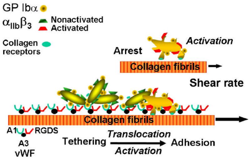
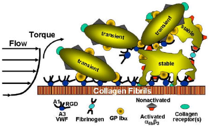
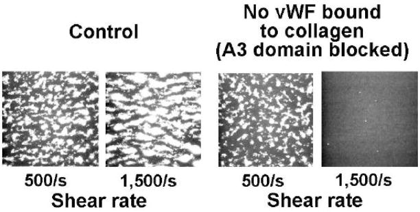
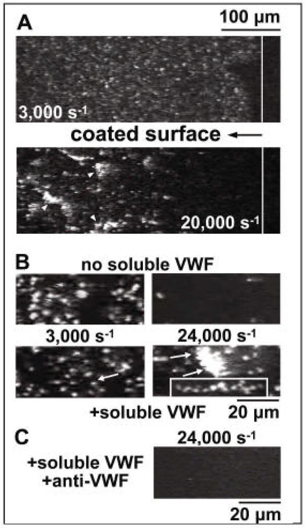
Von Willebrand factor (VWF) is a large multimeric glycoprotein produced in endothelial cells and megakaryocytes and present in subendothelial matrix, blood plasma and platelets. VWF mediates adhesion and aggregation of platelets at sites of vascular injury, processes that are critical for both haemostasis and thrombosis. Thrombus formation involves complex events that are influenced by different environmental conditions. Progress in understanding the structure and function of VWF and the mechanisms that underlie its interactions with platelets has led to important insight into the differentiation between normal haemostasis and pathological arterial thrombosis. The conventional view of signalling-induced platelet aggregation has recently been extended to include activation-independent aggregation. A novel mechanism has been demonstrated for initiating thrombus formation under high haemodynamic forces that involves alpha(IIb)beta(3)-independent platelet aggregation at the interface between immobilised and soluble VWF. This VWF-mediated process may be a key determinant of platelet accumulation in stenotic arteries leading to acute thrombotic occlusion.


Conflict of interest statement

Conflict of interest

None declared.

Figures

Figure 1
Figure 1
Schematic representation of the mechanisms of platelet adhesion in flowing blood. At the time of vascular injury, particularly in vessels of high shear stress, the platelets initially adhere transiently to subendothelial von Willebrand factor (VWF) through the glycoprotein (GP) Ib receptor. This contact significantly slows the movement of the platelets and leads to transient arrests, which then allow the engagement of other adhesive receptors, such as the collagen receptors shown here, resulting in a stable attachment. Modified from [4].
Figure 2
Figure 2
Schematic representation of the mechanisms of platelet aggregation in flowing blood. After stable adhesion, platelets are activated, secrete the contents of their granules and bind adhesive proteins from plasma, such as fibrinogen and von Willebrand factor (VWF), which form the substrate onto which additional platelets are recruited to form a thrombus. Modified from [10].
Figure 3
Figure 3
Single frames from a real-time recording of perfusion studies demonstrating how inhibiting von Willebrand factor (VWF) binding to collagen using a monoclonal antibody against the VWF A3 domain obliterates thrombus formation when the wall shear rate is 1500 s−1 but not 500 s−1. Adapted from [10].
Figure 4
Figure 4
Activation-independent platelet aggregation at the interface of immobilised and soluble VWF. A. Blood containing PPACK (anticoagulant), mepacrine (fluorescent dye), prostaglandin E1 (platelet inactivator) and EDTA (to prevent ligand binding to integrins) was perfused over immobilised von Willebrand factor (VWF). The vertical solid white line distinguishes VWF-coated (left) from uncoated (right) glass. Upper A panel illustrates single platelet adhesion at a shear rate of 3,000 s−1. Lower A panel illustrates rolling aggregates at a shear rate of 20,000 s−1. B. Perfusion over immobilised VWF of washed blood cells. Upper B panels illustrate the results in the absence of added purified VWF, i.e. efficient single platelet adhesion at a shear rate of 3,000 s−1 (left) and adhesion of only a few single platelets at 24,000 s−1 (right). Lower B panels illustrate the results after addition of purified VWF, i.e. single platelet adhesion at a shear rate of 3,000 s−1 (left), but formation of adhering aggregates at 24,000/s (right). The lower right-hand box of panel B illustrates a stretched aggregate during stationary adhesion. C. Perfusion over immobilised VWF of washed blood cells in the presence of added soluble VWF and anti-VWF A1 domain monoclonal antibody [11]. This research was originally published in Blood. Ruggeri ZM, Orje JN, Habermann R, Federici AB, Reininger AJ. Activation-independent platelet adhesion and aggregation under elevated shear stress. Blood 2006;108:1903–10. © the American Society of Hematology.

Similar articles

Cited by

References

    1. Titani K, Kumar S, Takio K, Ericsson LH, Wade RD, Ashida K, et al. Amino acid sequence of human von Willebrand factor. Biochemistry. 1986;25:3171–84. - PubMed
    1. Mendolicchio GL, Ruggeri ZM. New perspectives on von Willebrand factor functions in hemostasis and thrombosis. Semin Hematol. 2005;42:5–14. - PubMed
    1. Ruggeri ZM. Structure and function of von Willebrand factor. Thromb Haemost. 1999;82:576–84. - PubMed
    1. Wagner DD, Olmsted JB, Marder VJ. Immunolocalization of von Willebrand protein in Weibel-Palade bodies of human endothelial cells. J Cell Biol. 1982;95:355–60. - PMC - PubMed
    1. Bowie EJ, Solberg LA, Jr, Fass DN, Johnson CM, Knutson GJ, Stewart ML, et al. Transplantation of normal bone marrow into a pig with severe von Willebrand’s disease. J Clin Invest. 1986;78:26–30. - PMC - PubMed

Publication types

MeSH terms

Substances