

CD8+ T cells directed against a viral peptide contribute to loss of motor function by disrupting axonal transport in a viral model of fulminant demyelination
- PMID: 17493690
- PMCID: PMC1986839
- DOI: 10.1016/j.jneuroim.2007.04.005
CD8+ T cells directed against a viral peptide contribute to loss of motor function by disrupting axonal transport in a viral model of fulminant demyelination
Abstract
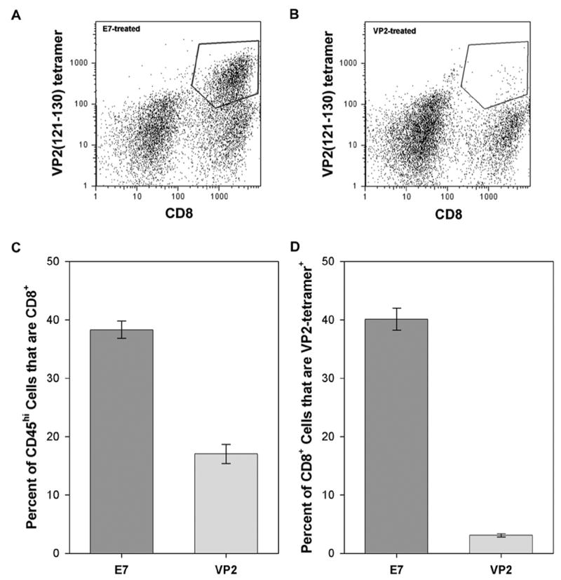
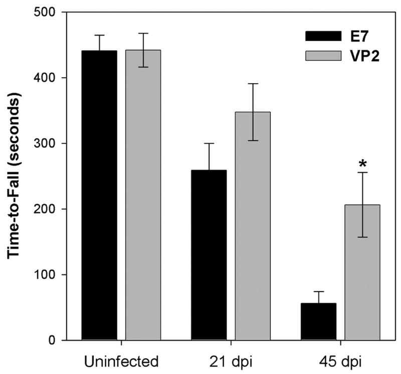
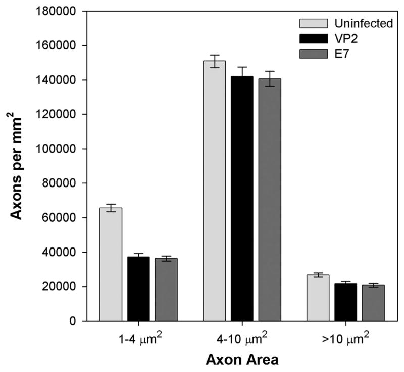
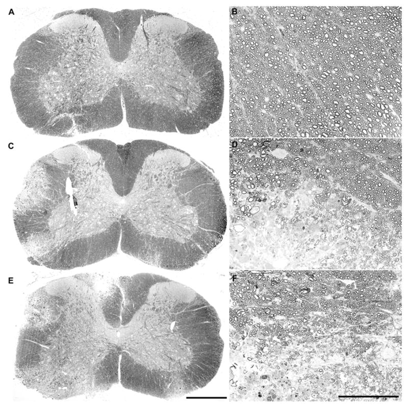
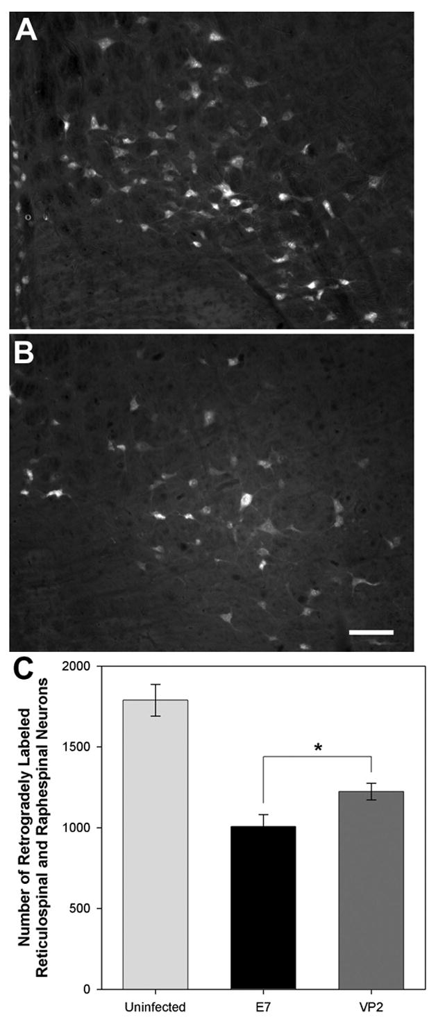
Demyelination, a pathological hallmark of multiple sclerosis, may be a necessary but not a sufficient condition for motor dysfunction associated with this disease. We favor a neurodegenerative model of multiple sclerosis and suggest that demyelination creates a permissive environment wherein the denuded axon becomes susceptible to immune-mediated injury. Unfortunately, the cellular effectors responsible for eliciting such axonal injury are currently unknown. Based on previous observations implicating cytotoxic T cells in this injury, we assessed motor function, axon dropout, and axon injury following peptide depletion of the immunodominant CD8+ antiviral T cell response in the IFNgamma receptor-deficient mouse model of acute demyelination. We found that the targeted removal of this population of cytotoxic effector cells prior to infection with the Theiler's murine encephalomyelitis virus caused a substantial preservation of motor function at 45 days postinfection that was associated with preservation of retrograde axonal transport in a subpopulation of surviving axons within the spinal cord. We conclude that cytotoxic T cells may be responsible for the initiation of axon injury following demyelination.
Figures
Similar articles
-
Preservation of motor function by inhibition of CD8+ virus peptide-specific T cells in Theiler's virus infection.FASEB J. 2001 Dec;15(14):2760-2. doi: 10.1096/fj.01-0373fje. Epub 2001 Oct 15. FASEB J. 2001. PMID: 11606479
-
Demyelinated axons and motor function are protected by genetic deletion of perforin in a mouse model of multiple sclerosis.J Neuropathol Exp Neurol. 2009 Sep;68(9):1037-48. doi: 10.1097/NEN.0b013e3181b5417e. J Neuropathol Exp Neurol. 2009. PMID: 19680139 Free PMC article.
-
Cellular sources and targets of IFN-gamma-mediated protection against viral demyelination and neurological deficits.Eur J Immunol. 2002 Mar;32(3):606-15. doi: 10.1002/1521-4141(200203)32:3<606::AID-IMMU606>3.0.CO;2-D. Eur J Immunol. 2002. PMID: 11857334 Free PMC article.
-
Inside-Out versus Outside-In models for virus induced demyelination: axonal damage triggering demyelination.Springer Semin Immunopathol. 2002;24(2):105-25. doi: 10.1007/s00281-002-0105-z. Springer Semin Immunopathol. 2002. PMID: 12503060 Free PMC article. Review.
-
Neuropathogenesis of Theiler's murine encephalomyelitis virus infection, an animal model for multiple sclerosis.J Neuroimmune Pharmacol. 2010 Sep;5(3):355-69. doi: 10.1007/s11481-009-9179-x. Epub 2009 Nov 6. J Neuroimmune Pharmacol. 2010. PMID: 19894121 Free PMC article. Review.
Cited by
-
CD8+ T cells cause disability and axon loss in a mouse model of multiple sclerosis.PLoS One. 2010 Aug 30;5(8):e12478. doi: 10.1371/journal.pone.0012478. PLoS One. 2010. PMID: 20814579 Free PMC article.
-
Isolation of brain-infiltrating leukocytes.J Vis Exp. 2011 Jun 13;(52):2747. doi: 10.3791/2747. J Vis Exp. 2011. PMID: 21694694 Free PMC article.
-
The CD8 T Cell-Epstein-Barr Virus-B Cell Trialogue: A Central Issue in Multiple Sclerosis Pathogenesis.Front Immunol. 2021 Jul 7;12:665718. doi: 10.3389/fimmu.2021.665718. eCollection 2021. Front Immunol. 2021. PMID: 34305896 Free PMC article. Review.
-
Molecular Mechanisms in the Genesis of Seizures and Epilepsy Associated With Viral Infection.Front Mol Neurosci. 2022 May 9;15:870868. doi: 10.3389/fnmol.2022.870868. eCollection 2022. Front Mol Neurosci. 2022. PMID: 35615063 Free PMC article. Review.
-
Axonopathy is associated with complex axonal transport defects in a model of multiple sclerosis.Brain Pathol. 2012 Jul;22(4):454-71. doi: 10.1111/j.1750-3639.2011.00541.x. Epub 2011 Nov 17. Brain Pathol. 2012. PMID: 21988534 Free PMC article.
References
-
- Babbe H, Roers A, Waisman A, Lassmann H, Goebels N, Hohlfeld R, Friese M, Schroder R, Deckert M, Schmidt S, Ravid R, Rajewsky K. Clonal expansions of CD8(+) T cells dominate the T cell infiltrate in active multiple sclerosis lesions as shown by micromanipulation and single cell polymerase chain reaction. J Exp Med. 2000;192:393–404. - PMC - PubMed
-
- Bitsch A, Schuchardt J, Bunkowski S, Kuhlmann T, Bruck W. Acute axonal injury in multiple sclerosis. Correlation with demyelination and inflammation. Brain. 2000;123 ( Pt 6):1174–1183. - PubMed
-
- Buenz EJ, Howe CL. Picornaviruses and cell death. Trends Microbiol. 2006;14:28–36. - PubMed
-
- Compston A. The pathogenesis and basis for treatment in multiple sclerosis. Clin Neurol Neurosurg. 2004;106:246–248. - PubMed
Publication types
MeSH terms
Substances
Grants and funding
LinkOut - more resources
Full Text Sources
Molecular Biology Databases
Research Materials
