. 2007 Jul;98(1):105-21.
doi: 10.1152/jn.00330.2007. Epub 2007 May 9.

Dynamic circuitry for updating spatial representations. III. From neurons to behavior

Affiliations

Dynamic circuitry for updating spatial representations. III. From neurons to behavior

Rebecca A Berman et al. J Neurophysiol. 2007 Jul.

Abstract

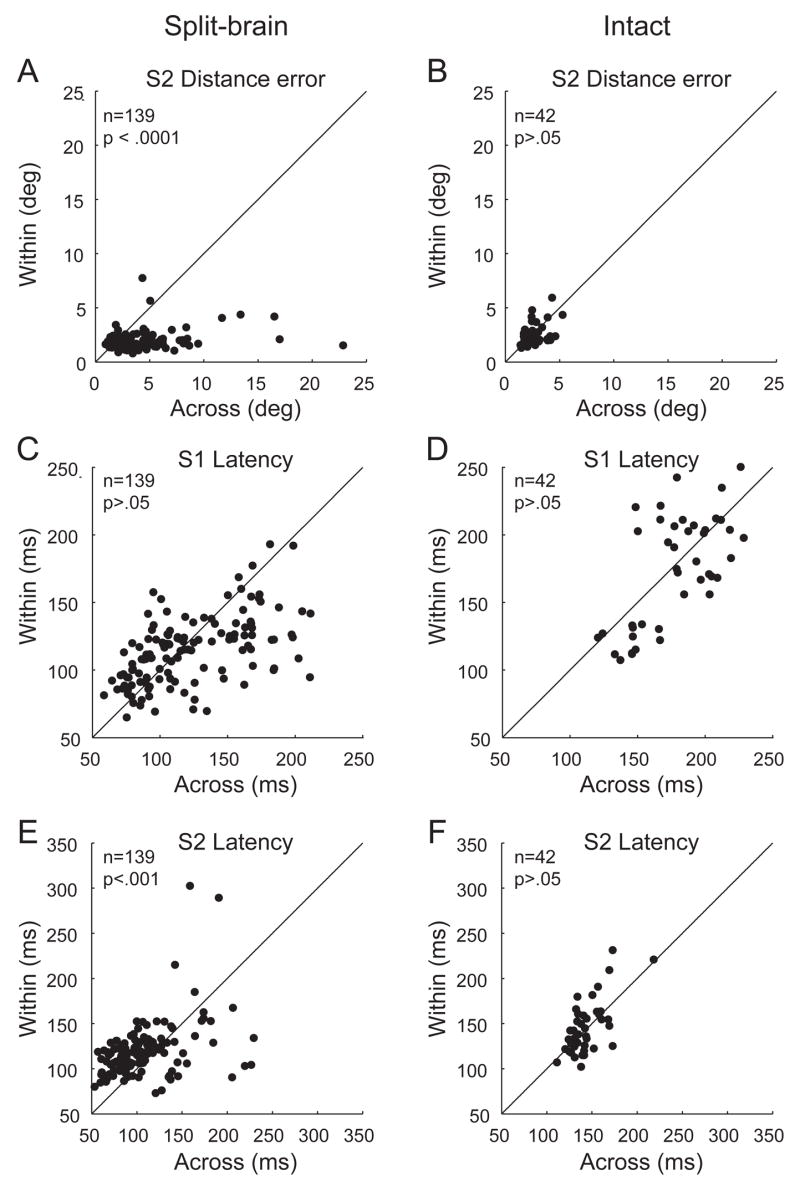
Each time the eyes move, the visual system must adjust internal representations to account for the accompanying shift in the retinal image. In the lateral intraparietal cortex (LIP), neurons update the spatial representations of salient stimuli when the eyes move. In previous experiments, we found that split-brain monkeys were impaired on double-step saccade sequences that required updating across visual hemifields, as compared to within hemifield. Here we describe a subsequent experiment to characterize the relationship between behavioral performance and neural activity in LIP in the split-brain monkey. We recorded from single LIP neurons while split-brain and intact monkeys performed two conditions of the double-step saccade task: one required across-hemifield updating and the other required within-hemifield updating. We found that, despite extensive experience with the task, the split-brain monkeys were significantly more accurate for within-hemifield than for across-hemifield sequences. In parallel, we found that population activity in LIP of the split-brain monkeys was significantly stronger for the within-hemifield than for the across-hemifield condition of the double-step task. In contrast, in the normal monkey, both the average behavioral performance and population activity showed no bias toward the within-hemifield condition. Finally, we found that the difference between within-hemifield and across-hemifield performance in the split-brain monkeys was reflected at the level of single-neuron activity in LIP. These findings indicate that remapping activity in area LIP is present in the split-brain monkey for the double-step task and covaries with spatial behavior on within-hemifield compared to across-hemifield sequences.


Figures

Fig. 1
Fig. 1
Assessment of behavior and neural activity during within- and across-hemifield remapping in the split-brain monkey. The geometry of the double-step sequence is determined by the location of the neuron’s receptive field. The hypothetical neuron under study is located in the left hemisphere (grey asterisk), with a receptive field (circle) in the upper right visual field. In the across-hemifield condition (A), the second target (T2) is located in the left visual field when the eyes are at central fixation (FP); its location is represented by neurons in the right hemisphere (black asterisk). When the eyes reach the first target (T1), however, the location where T2 appeared is now in the right visual field; its stimulus trace is thus represented by neurons in the left hemisphere, including the neuron under study (gray asterisk). Updating in this condition involves a transfer of visual information between neurons in opposite cortical hemispheres. In the within-hemifield condition (B) T2 appears in the right visual field when the eyes are at FP, and so is represented by neurons in the left hemisphere (black asterisk). After the saccade to T1, the stimulus trace of T2 is still represented by neurons in the left hemisphere (gray asterisk). Updating in this condition therefore involves the transfer of visual signals within the same hemisphere.
Fig. 2
Fig. 2
Activity in a single neuron that remaps both within and across hemifields in the double-step task. The monkey makes sequential saccades from fixation (crosshair) to the first target (dot) and second target (asterisk). The first saccade brings the neuron’s receptive field onto the location where the second target had appeared. Eye position traces from the first ten trials are shown for the within (A) and across (B) conditions. Recording during the double-step task shows neural activity during the within (C) and across (D) conditions. Recording during the single-step task likewise shows that activity in both the within (E) and across (F) conditions is greater than in the control tasks (G–J).
Fig. 3
Fig. 3
Accuracy and latency of double-step performance. Panels in the left column represent data from the two split-brain monkeys; those in the right column are from a monkey with commissures intact. Each point represents average double-step behavior during the recording of a single neuron, for the within-hemifield (y axis) and across-hemifield condition (x axis). Distance error of the second saccade (S2) was significantly greater for across-hemifield updating in the split-brain (A) but not the intact animal (B). For latency (C–F), both the first and second saccades were faster overall for the split-brain monkeys, who were highly experienced on the task as compared to the intact monkey. For the first saccade (S1), within-hemifield and across-hemifield latencies were not significantly different for either split-brain or intact (C,D). For the second saccade, overall latencies were significantly faster for the across-hemifield condition in the split-brain (E) but did not differ in the intact monkey (F).
Fig. 4
Fig. 4
Relationship between speed of saccade initiation and accuracy for the second saccade of the double-step task. Each point represents the spatial error (y axis) and average saccade latency (x axis) from a given neural recording session. Each session contributes two datapoints, one from within-hemifield trials and one from across-hemifield trials. The regression line is indicated by the thin black line. For both the split-brain (A) and intact (B) monkey, the overall slope of the relationship between speed and accuracy is positive: as latency increased, so did spatial error.
Fig. 5
Fig. 5
In the split-brain monkey, updating activity in LIP is stronger for the within-hemifield than the across-hemifield condition. This difference was not observed in the intact monkey. Bars represent the percentage of neurons with significant remapping for Within only, Across only, or both conditions in the split-brain (A) and intact monkey (B); neurons with no significant remapping are excluded. For all single neurons in the split-brain (C) and intact monkey (D), average updating activity in the within-hemifield condition (y axis) is plotted against that in the across-hemifield condition (x axis). Updating activity is adjusted to take saccade-alone activity into account (see Methods); negative updating activity (below or to the left of the dotted lines in C,D) indicates that saccade-alone activity exceeded activity in the double-step task.
Fig. 6
Fig. 6
In the split-brain monkey (A), neural activity begins earlier for within-hemifield than across-hemifield updating. This difference was not observed in the intact monkey (B). Analysis was conducted on a subset of neurons with detectable neural latencies for both within and across conditions (see RESULTS). For each neuron, the neural latency in the within-hemifield condition (y axis) is plotted against that in the across-hemifield condition (x axis). Neural latency is determined relative to the beginning of the first saccade (S1). Grey shading represents the region in which both within-hemifield and across-hemifield neural latencies were after the start of S1; unshaded areas indicate neural activity that began even before the start of S1. This activity reflects predictive updating, which was more frequent in the split-brain monkey for the within than the across condition. This is consistent with delayed across-hemifield updating activity in the absence of the forebrain commissures.
Fig. 7
Fig. 7
Comparison of neural activity in the single-step and double-step tasks. For the split-brain (A) and intact animal (B), average firing rate is greater in the double-step as compared to the single-step task. Each dot represents the average firing rate for a single neuron for the double-step task (y axis) plotted against that for the single-step task (x axis). Each neuron contributes two data points, one for within and one for across. For the split-brain (C) and intact monkey (D), increased activity in the double-step task is present for both within-hemifield and across-hemifield updating. Each point represents the differential activity for the two tasks (Double-step minus Single-step, in sp/s). The average differential activity for each neuron is shown for the within condition (y axis) and across condition (x axis). The shaded square shows the region in which double-step activity is greater than single-step activity for both within-and across- hemifield conditions.
Fig. 8
Fig. 8
Population measures of updating strength and behavioral accuracy and latency. Each panel shows the distribution of Within:Across (WA) index values from all neurons (single recording sessions). The index represents a bias for within-hemifield updating (positive values) or across-hemifield updating (negative values). The vertical line indicates no difference between within- and across-hemifield updating. In the split-brain monkey, the WA index is positively skewed for neural activity and distance error (A,C), but is negatively skewed for saccade latency (E). In the intact monkey, the WA index distribution does not differ from zero for neural activity (B), accuracy (D), or latency (F). Thus, at the population level, saccade accuracy parallels neural activity for both split-brain and intact monkeys. Saccade latency, by contrast, parallels neural activity in the intact monkey but not in the split-brain monkeys.
Fig. 9
Fig. 9
Trial-by-trial analysis for single-neuron updating activity in LIP and the accuracy of double-step performance. Panels show the distribution of r values for the trial-by-trial correlation between updating activity and error of the second saccade. The left column shows data from the split-brain monkeys, right column from the intact monkey. Top row (A,B) shows data for all trials (within and across combined), middle row (C,D) shows data for within-hemifield trials only, and bottom row for across-hemifield trials only (E,F). Black shading indicates single neurons for which the trial-by-trial relationship was significant at p < .05. Vertical line indicates zero; x axis is identical for all panels. Negative r values indicate that greater updating activity was associated with smaller error. In the split-brain monkey, the population of r values had a significant negative skew only when all trials were combined (A), due to the differences between within and across-hemifield updating. The distributions were not significantly skewed for separate within (C) or across (E) trials in the split-brain monkey. In the intact monkey, there was no relationship between updating activity and second-saccade error on the double-step task whether trials were combined (B) or separate (D,F).
Fig. 10
Fig. 10
Trial-by-trial analysis for single-neuron updating activity in LIP and the latency of double-step performance. Panels show the distribution of r values for the trial-by-trial correlation between updating activity and latency of the second saccade. Conventions as in Fig. 9. For all trials combined (A,B) the distribution had a slight but nonsignificant positive skew for both split-brain and intact monkey, indicating that stronger updating activity was associated with longer latencies. This positive skew was significant in the intact monkey for within-hemifield trials alone (D). For all other cases in the split-brain (A,C,E) and intact monkey (B,F), there was no significant trial-by-trial relationship between updating activity and second-saccade latency on the double-step task.
Fig. 11
Fig. 11
Changes in behavior and neural activity over multiple sessions of testing with the same spatial geometry of the double-step saccade task. Each point represents data from a single recording session (one cell) in which the receptive field (RF) was located at the same angle, 45°, from fixation (A). In panels B–D, Within:Across indices (y axis) are plotted as a function of session (x axis); regression lines are indicated by the thin black lines. Positive index values denote greater accuracy (B), faster latency (C), or stronger updating activity (D) for the within-hemifield sequence. For accuracy and updating activity, index values approached zero over sessions, indicating that within and across updating became more alike as the monkey gained experience. For latency, index values became increasingly more negative, indicating that across-hemifield latencies became even shorter relative to within-hemifield latencies as the monkey gained experience.

References

    1. Andersen RA. Handbook of Physiology: The Nervous System. Bethesda, MD: Williams Wekens; 1987. Inferior parietal lobule function in spatial perception and visuomotor integration; pp. 483–518.
    1. Assad JA. Neural coding of behavioral relevance in parietal cortex. Curr Opin Neurobiol. 2003;13:194–197. - PubMed
    1. Baizer JS, Bender DB. Comparison of saccadic eye movements in humans and macaques to single-step and double-step target movements. Vision Res. 1989;29:485–495. - PubMed
    1. Baker JT, Harper TM, Snyder LH. Spatial memory following shifts of gaze. I. Saccades to memorized world-fixed and gaze-fixed targets. J Neurophysiol. 2003;89:2564–2576. - PubMed
    1. Barash S, Bracewell RM, Fogassi L, Gnadt JW, Andersen RA. Saccade-related activity in the lateral intraparietal area. I. Temporal properties; comparison with area 7a. J Neurophysiol. 1991;66:1095–1108. - PubMed

Publication types

LinkOut - more resources