. 2007 Jul;98(1):334-44.
doi: 10.1152/jn.00202.2007. Epub 2007 May 9.

In vivo roles for matrix metalloproteinase-9 in mature hippocampal synaptic physiology and plasticity

Affiliations

In vivo roles for matrix metalloproteinase-9 in mature hippocampal synaptic physiology and plasticity

Ozlem Bozdagi et al. J Neurophysiol. 2007 Jul.

Abstract

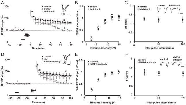
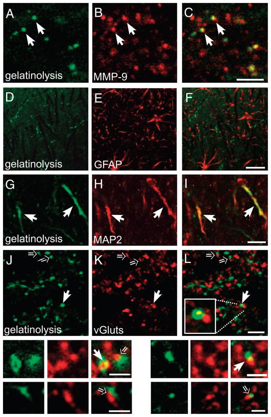
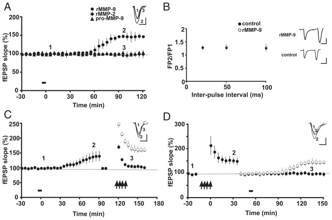
Extracellular proteolysis is an important regulatory nexus for coordinating synaptic functional and structural plasticity, but the identity of such proteases is incompletely understood. Matrix metalloproteinases (MMPs) have well-known, mostly deleterious roles in remodeling after injury or stroke, but their role in nonpathological synaptic plasticity and function in intact adult brains has not been extensively investigated. Here we address the role of MMP-9 in hippocampal synaptic plasticity using both gain- and loss-of-function approaches in urethane-anesthetized adult rats. Acute blockade of MMP-9 proteolytic activity with inhibitors or neutralizing antibodies impairs maintenance, but not induction, of long-term potentiation (LTP) at synapses formed between Schaffer-collaterals and area CA1 dendrites. LTP is associated with significant increases in levels of MMP-9 and proteolytic activity within the potentiated neuropil. By introducing a novel application of gelatin-substrate zymography in vivo, we find that LTP is associated with significantly elevated numbers of gelatinolytic puncta in the potentiated neuropil that codistribute with immunolabeling for MMP-9 and for markers of synapses and dendrites. Such increases in proteolytic activity require NMDA receptor activation. Exposing intact area CA1 neurons to recombinant-active MMP-9 induces a slow synaptic potentiation that mutually occludes, and is occluded by, tetanically evoked potentiation. Taken together, our data reveal novel roles for MMP-mediated proteolysis in regulating nonpathological synaptic function and plasticity in mature hippocampus.


Figures

FIG. 1
FIG. 1
Matrix metalloproteinase 9 (MMP-9) inhibition impairs long-term potentiation (LTP) maintenance in adult rat area CA1 in vivo. A: tetanic stimulation (↑, 4 trains of 100 Hz, 1-s stimulation separated by 5 min) of the Schaffer collaterals in a control group produces robust potentiation of field excitatory postsynaptic potentials (fEPSPs; ●). In contrast, in rats subjected to intrahippocampal injection of the MMP-9 blocker Inhibitor II (250 μM; bar), potentiation returns to baseline by 90–120 min posttetanic stimulation (○) with no significant effects on LTP induction through the 1st 30 min (P > 0.3 at 30 min compared with control values). There were no effects on LTP after injection of the diluent alone (0.6% DMSO; ▵). Inset: representative EPSP traces were recorded before tetanus (1), 120 min after tetanus in the presence of Inhibitor II (2) and 120 min after tetanus in the presence of the DMSO diluent (3). Calibration: 10 ms, 0.3 mV. B: input–output curves, representing the relationship between stimulus intensity and the size of the fEPSP slope, did not differ significantly between animals injected with Inhibitor II (250 μM, ○) and control animals (●). C: there were no differences in the paired-pulse facilitation (PPF) ratios between animals injected with Inhibitor II (250 μM) and control animals. The PPF ratio represents the slope of the second fEPSP (FP2) divided by the slope of the first fEPSP (FP1) for the interpulse intervals (IPI) shown. Inset: representative EPSP traces from control and inhibitor-treated animals (IPI: 50 ms). Calibration: 25 ms, 0.2 mV. D: rats receiving an intrahippocampal injection of an MMP-9 function-blocking antibody (15 min, bar) show a deficit in LTP maintenance (○) in comparison with control animals (●). Such deficits were similar to those observed with Inhibitor II shown in A. There were no effects on LTP in animals receiving an intrahippocampal injection of nonimmune mouse IgG (▵). Inset: representative EPSP traces were recorded before tetanus (1), 120 min after tetanus in the presence of MMP-9 function-blocking antibody (2) and 120 min after tetanus in the presence of IgG control antibody (3). Calibration as in A. E: there were no differences between control animals and those receiving MMP-9 blocking antibody in input–output curves. F: there were no differences in the PPF ratios between animals injected with MMP-9 blocking antibody and control animals. Inset: representative EPSP traces from control and MMP-9 antibody-treated animals. Other conventions as in C.
FIG. 2
FIG. 2
Regulation of MMP-9 levels and proteolytic activity during LTP. A: representative immunoblots (left) of rat area CA1 homogenates (n = 3 rats) frozen 60 min post-LTP induction. For each animal, homogenates were prepared from ipsilateral (LTP) and contralateral (contra) sides. Membranes were probed with antibodies that recognize both the pro- and active-form of MMP-9 (pro-9, act-9 respectively) or glyceraldehyde-3-phosphate dehydrogenase (GAPDH), used as a loading control. Quantification of band intensity (right) showed significant increases in levels of both pro- and active-forms of MMP-9 in sides undergoing LTP in comparison with those in contralateral sides (*P < 0.05). B: low-magnification confocal image of rat hippocampus showing representative injection of the MMP-9 substrate DQ gelatin into area CA1. SO, stratum oriens; P, s. pyramidale; SR, s. radiatum; S L-M, s. lacunosummoleculare. Bar, 500 μm. C: intrahippocampal injection of DQ-gelatin (bar) has no effects on induction or maintenance of LTP (○, n = 7) in comparison with control animals (●; n = 6, P > 0.05 at 60 min). LTP is abolished in animals receiving an intraperitoneal injection of the N-methyl-D-aspartate (NMDA) receptor antagonist 3-(2-carboxypiperazin-4-yl)propyl-1-phosphonic acid (CPP) prior to tetanic stimulation (triangles, n = 3). Inset: representative EPSP traces were recorded before and 60 min after LTP induction in control, DQ-gelatin (gel) injected and CPP-administered animals. Calibration: 10 ms, 0.5 mV. D—F: high-magnification confocal images through area CA1 s. radiatum taken from animals 75 min after receiving control stimulation (D), LTP-inducing tetanic stimulation (E), or LTP-inducing tetanic stimulation after CPP administration (F). Numerous “hot spots” of gelatinolytic puncta are evident within the potentiated neuropil in the LTP animals in comparison with very few puncta present in animals receiving control stimuli or those in which LTP was blocked by CPP. Bars, 10 μm. G: quantitative analysis of numbers of gelatinolytic puncta within area CA1 s. radiatum. There was significantly greater numbers of puncta in LTP animals in comparison with the other 2 groups (*P < 0.01).
FIG. 3
FIG. 3
Gelatinolytic puncta codistribute with dendritic and synaptic markers. A–L, insets: high-power confocal images through area CA1 s. radiatum taken 75 min after LTP induction in rats receiving an intrahippocampal injection of the MMP-9 substrate DQ-gelatin. Left: gelatinolytic puncta (green); middle: immunolabeling for indicated markers (red); and right: merged images; regions of codistributed overlap are indicated by yellow pixels. A–C: most gelatinolytic puncta codistribute with immunolabeling for MMP-9 (arrows). There are numerous MMP-9 immunolabeled puncta that are not gelatinolytic, which presumably represents a pool of inactive (pro) MMP-9, as the antibody recognizes both pro- and active-forms of MMP-9. D—F: gelatinolytic puncta did not codistribute with GFAP immunolabeling, a marker of astrocytes. G–I: many puncta or elongated regions of gelatinolysis codistributed with immunolabeling for MAP-2, a dendritic marker (arrows). J–L: many gelatinolytic puncta codistributed with immunolabeling for vGluts, a presynaptic terminal marker (solid arrows), whereas others appeared abutted or directly apposed to vGlut-immunolabeled puncta (open arrows). Insets: underneath show higher-power images of such labeling patterns. Bars, 3 μm A-L; 2 μm in insets.
FIG. 4
FIG. 4
There are no changes in MMP-9 mRNA levels or distribution with LTP. A and B: representative film autoradiograms of bilateral hipppocampal sections showing pattern and relative intensity of MMP-9 cRNA probe hybridization. Section in A was taken from an animal 75 min after receiving control stimuli on one side (control stim); section in B was taken from an animal 75 min after LTP was induced tetanically on 1 side (LTP). In both images, the contralateral sides (contra) are also shown for comparison. There are no apparent differences in levels or patterns of probe hybridization across sides or stimulation conditions. Bars, 2 mm. C: quantitative densitometry shows no differences in relative levels of probe hybridization between LTP (n = 6) animals and those receiving control stimuli (n = 4) in s. pyramidale of areas CA1 and CA3, the granule cell layer of the dentate gyrus (DG), or area CA1 st. radiatum (st. rad).
FIG. 5
FIG. 5
Proteolytically active MMP-9 induces potentiation in rat area CA1 in vivo that occludes, and is occluded by, tetanic stimulation. A: recombinant active MMP-9 (rMMP-9; 0.1 μg/μl) delivered intrahippocampally (bar) induces a slowly emerging, persistent potentiation that begins to arise approximately 60 min postinjection (●, n = 5). Administration of inactive pro-MMP-9 (▲) or recombinant active MMP-2 (rMMP-2; ◆) did not elicit an increase in fEPSP slope (n = 3 each). Inset: representative EPSP traces were recorded before (1), 90 min after rMMP-9 application (2), and 90 min after rMMP-2 application (3). Calibration: 10 ms, 0.3 mV. B: intrahippocampal infusion of rMMP-9 has no effect on PPF. Plot shows PPF measured before (●) and 120 min after (○) rMMP-9 administration. Inset: representative EPSP traces (50 ms IPI) recorded before and after rMMP-9 injection. Calibration: 25 ms, 0.2 mV. C: rMMP-9 potentiation occludes tetanically-induced potentiation. After rMMP-9 potentiation reached a plateau (~90 min), fEPSP slope was reduced to yield synaptic responses that matched those of baseline levels, followed by tetanic stimulation (↑). There was no further potentation after tetanic stimulation (●). The ghostline (○) indicates the level of tetantically induced LTP in control animals (see Fig. 1A). Representative EPSP traces were recorded at time points indicated in the panel. D: tetanic stimulation occludes rMMP-9 potentiation. Tetanic stimulation (↑) produces LTP (●); at ~45 min postinduction, stimulus intensity was reduced to produce synaptic responses that matched those of the original baseline, followed by administration of rMMP-9 (bar). There was no further potentiation following delivery of rMMP-9 (●). The ghostline (○) indicates the level of rMMP-9-induced potentiation from animals shown in A. Representative EPSP traces were recorded at time points indicated in the panel. Calibration as in Fig. 1A.

Similar articles

Cited by

References

    1. Antonova I, Arancio O, Trillat AC, Wang HG, Zablow L, Udo H, Kandel ER, Hawkins RD. Rapid increase in clusters of presynaptic proteins at onset of long-lasting potentiation. Science. 2001;294:1547–1550. - PubMed
    1. Arai K, Lee SR, Lo EH. Essential role for ERK mitogen-activated protein kinase in matrix metalloproteinase-9 regulation in rat cortical astrocytes. Glia. 2003;43:254–264. - PubMed
    1. Bahr BA, Staubli U, Xiao P, Chun D, Ji Z-X, Esteban ET, Lynch G. Arg-gly-asp-ser-selective adhesion and the stabilization of long-term potentiation: pharmacological studies and the characterization of a candidate matrix receptor. J Neurosci. 1997;17:1320–1329. - PMC - PubMed
    1. Baranes D, Lederfein D, Huang YY, Chen M, Bailey CH, Kandel ER. Tissue plasminogen activator contributes to the late phase of LTP and to synaptic growth in the hippocampal mossy fiber pathway. Neuron. 1998;21:813–825. - PubMed
    1. Barlow JZ, Kelley KA, Bozdagi O, Huntley GW. Testing the role of the cell-surface molecule Thy-1 in regeneration and plasticity of connectivity in the CNS. Neuroscience. 2002;111:837–852. - PubMed

Publication types

MeSH terms

Substances

LinkOut - more resources