. 2007 Jul;81(14):7776-85.
doi: 10.1128/JVI.00546-06. Epub 2007 May 9.

Induction of MxA gene expression by influenza A virus requires type I or type III interferon signaling

Affiliations

Induction of MxA gene expression by influenza A virus requires type I or type III interferon signaling

Dirk Holzinger et al. J Virol. 2007 Jul.

Abstract

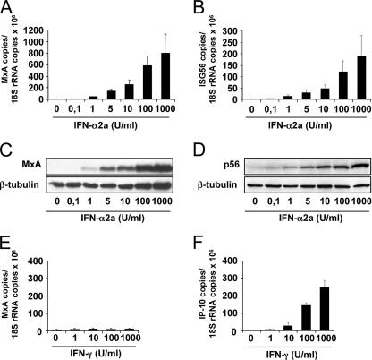
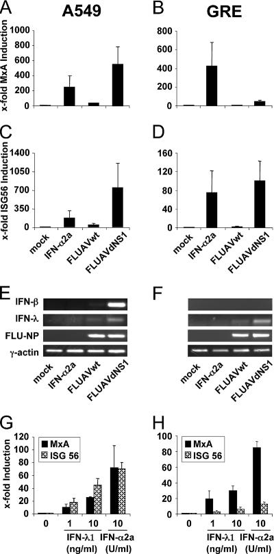
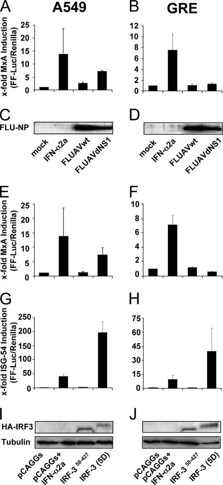
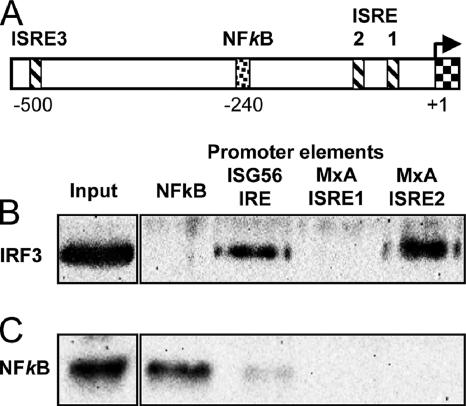
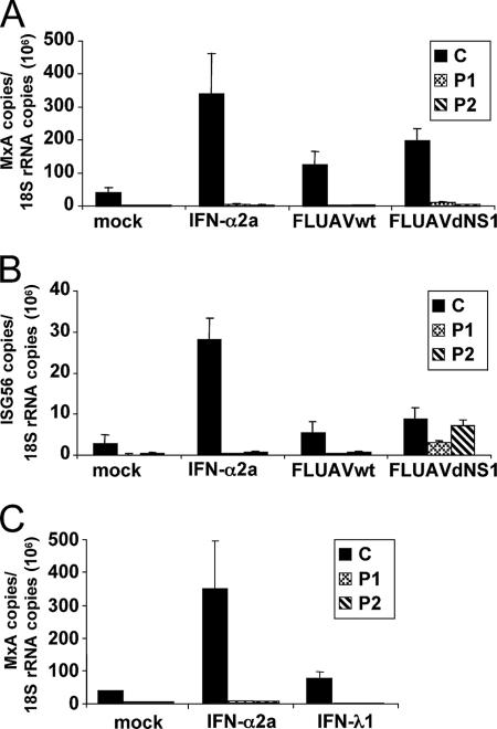
The human MxA gene belongs to the class of interferon (IFN)-stimulated genes (ISGs) involved in antiviral resistance against influenza viruses. Here, we studied the requirements for MxA induction by influenza A virus infection. MxA is transcriptionally upregulated by type I (alpha and beta) and type III (lambda) IFNs. Therefore, MxA is widely used in gene expression studies as a reliable marker for IFN bioactivity. It is not known, however, whether viruses can directly activate MxA expression in the absence of secreted IFN. By using an NS1-deficient influenza A virus and human cells with defects in IFN production or the STAT1 gene, we studied the induction profile of MxA by real-time reverse transcriptase PCR. The NS1-deficient virus is known to be a strong activator of the IFN system because NS1 acts as a viral IFN-antagonistic protein. Nevertheless, MxA gene expression was not inducible by this virus upon infection of IFN nonproducer cells and STAT1-null cells. Likewise, neither IFN-alpha nor IFN-lambda had a sizeable effect on the STAT1-null cells, indicating that MxA expression requires STAT1 signaling and cannot be triggered directly by virus infection. In contrast, the expression of the IFN-stimulated gene ISG56 was induced by influenza virus in these cells, confirming that ISG56 differs from MxA in being directly inducible by viral triggers in an IFN-independent way. In summary, our study reveals that MxA is a unique marker for the detection of type I and type III IFN activity during virus infections and IFN therapy.


Figures

FIG. 1.
FIG. 1.
Induction of MxA expression by IFN-α and IFN-γ. PBMCs from healthy donors (2 × 10⁶ cells) were treated with increasing concentrations of IFN-α2a (A to D) or IFN-γ (E and F). After 6 h, the amounts of MxA (A and E), ISG56 (B), and IP-10 (F) transcripts were determined by qRT-PCR and normalized to the amount of 18S rRNA transcripts. After 16 h, MxA protein (C) and p56 protein (D) were analyzed by Western blotting using specific antibodies. β-Tubulin was used as an internal standard. The error bars represent standard deviations calculated from three experiments.
FIG. 2.
FIG. 2.
MxA is induced by type I or type III IFNs in influenza A virus-infected cells. A549 (A, C, E, and G) and GRE (B, D, F, and H) cells were infected with 1 PFU per cell of either FLUAVwt or FLUAVdNS1. Parallel cultures were treated with 1,000 U/ml of IFN-α2a or were left untreated (mock). To monitor induction by IFN-λ (G and H), cells were incubated with IFN-λ1 (1 ng/ml or 10 ng/ml) or IFN-α2a (10 U/ml). After 16 h, the amounts of MxA (A, B, G, and H) and ISG56 (C, D, G, and H) were determined by qRT-PCR and normalized to the amount of HPRT transcripts. The induction in untreated cells was defined as onefold. Error bars represent standard deviations calculated from three experiments. The induction of IFN-β and IFN-λ (E and F) was monitored by conventional RT-PCR using specific primers. Transcripts of FLUAV-NP and γ-actin were analyzed to monitor virus infection and expression of a housekeeping gene, respectively (E and F).
FIG. 3.
FIG. 3.
MxA promoter activation in response to virus infection occurs via type I IFN. A549 and GRE cells were transfected with pMxAP-FF-Luc (A, B, E, and F) or pISG54-FF-Luc (G and H) reporter plasmids. As an internal control, the cells were cotransfected with the SV40-REN-Luc reporter plasmid. (A to D) Cells were infected 6 h later with 1 PFU per cell of FLUAVwt or FLUAVdNS1. Parallel cultures were treated with 1,000 U/ml IFN-α2a or were left untreated (mock). (E to J) Cells were cotransfected with pCAGGS or pCAGGS expression constructs coding for IRF3(58-427) or IRF3(5D). At 24 h postinfection (A to D) or posttransfection (E to J), cells were lysed and FF-Luc and REN-Luc activities were measured. The FF-Luc activities were normalized to that of REN-Luc. The induction in untreated cells was defined as onefold. Bars represent standard deviations calculated from three experiments. The expression of viral NP (C and D) or HA-tagged IRF3 (I and J) was monitored by Western blotting using NP- or HA-specific antibodies, respectively. β-Tubulin (I and J) was used as a loading control.
FIG. 4.
FIG. 4.
IRF3 binding to the MxA promoter. (A) Schematic presentation of the MxA promoter region (−1 to −530). The transcriptional start site is marked with an arrow (+1, exon 1). ISREs ISRE1 (−45 to −57), ISRE2 (−88 to −103), and ISRE3 (−490 to −505) are indicated with striped boxes, and one NF-κB site is indicated with a punctated box (−234 to −244) according to the data in reference . Binding of activated IRF3 (B) and NF-κB (C) to the Sepharose-immobilized NF-κB promoter element of IFN-β PRDII, IRE of the ISG56 promoter, or ISRE1 and ISRE2 of the MxA promoter. Nuclear extracts of FLUAVdNS1-infected A549 cells were incubated with the immobilized oligonucleotides, and DNA-bound proteins were detected by Western blot analysis using IRF3 and NF-κB p50-specific antibodies. The left panels show the respective protein signals in the nuclear extract. The results are representative of two individual experiments.
FIG. 5.
FIG. 5.
MxA induction depends on STAT1 signaling. Control cells, C, and two STAT1-deficient cell lines, P1 (STAT1/1757-1758delAG) and P2 (STAT1/L600P), were infected with 5 PFU per cell of FLUAVwt or FLUAVdNS1 or treated with 1,000 U/ml of IFN-α2a or left untreated. After 16 h, the amounts of MxA (A) and ISG56 (B) transcripts were determined by qRT-PCR and normalized to the amount of 18S rRNA transcripts. Error bars represent standard deviations calculated from three experiments. (C) Induction of MxA by IFN-λ. Cells were treated with 1,000 U/ml of IFN-α2a or 10 ng/ml IFN-λ1. The amounts of MxA and 18S rRNA transcripts were determined as described above.

Similar articles

Cited by

References

    1. Aebi, M., J. Fäh, N. Hurt, C. E. Samuel, D. Thomis, L. Bazzigher, J. Pavlovic, O. Haller, and P. Staeheli. 1989. cDNA structures and regulation of two interferon-induced human Mx proteins. Mol. Cell. Biol. 9:5062-5072. - PMC - PubMed
    1. Alexopoulou, L., A. C. Holt, R. Medzhitov, and R. A. Flavell. 2001. Recognition of double-stranded RNA and activation of NF-κB by Toll-like receptor 3. Nature 413:732-738. - PubMed
    1. Antonelli, G., E. Simeoni, O. Turriziani, R. Tesoro, A. Redaelli, L. Roffi, L. Antonelli, M. Pistello, and F. Dianzani. 1999. Correlation of interferon-induced expression of MxA mRNA in peripheral blood mononuclear cells with the response of patients with chronic active hepatitis C to IFN-α therapy. J. Interferon Cytokine Res. 19:243-251. - PubMed
    1. Asahina, Y., N. Izumi, M. Uchihara, O. Noguchi, Y. Nishimura, K. Inoue, K. Ueda, K. Tsuchiya, K. Hamano, J. Itakura, and S. Miyake. 2003. Interferon-stimulated gene expression and hepatitis C viral dynamics during different interferon regimens. J. Hepatol. 39:421-427. - PubMed
    1. Baechler, E. C., F. M. Batliwalla, G. Karypis, P. M. Gaffney, W. A. Ortmann, K. J. Espe, K. B. Shark, W. J. Grande, K. M. Hughes, V. Kapur, P. K. Gregersen, and T. W. Behrens. 2003. Interferon-inducible gene expression signature in peripheral blood cells of patients with severe lupus. Proc. Natl. Acad. Sci. USA 100:2610-2615. - PMC - PubMed

Publication types