

doi: 10.1128/JVI.00724-07.
Epub 2007 May 9.
Peptide-mediated interference with influenza A virus polymerase
Affiliations
- PMID: 17494067
- PMCID: PMC1933368
- DOI: 10.1128/JVI.00724-07
Item in Clipboard
Peptide-mediated interference with influenza A virus polymerase
J Virol.
2007 Jul.
Abstract
The assembly of the polymerase complex of influenza A virus from the three viral polymerase subunits PB1, PB2, and PA is required for viral RNA synthesis. We show that peptides which specifically bind to the protein-protein interaction domains in the subunits responsible for complex formation interfere with polymerase complex assembly and inhibit viral replication. Specifically, we provide evidence that a 25-amino-acid peptide corresponding to the PA-binding domain of PB1 blocks the polymerase activity of influenza A virus and inhibits viral spread. Targeting polymerase subunit interactions therefore provides a novel strategy to develop antiviral compounds against influenza A virus or other viruses.
Figures

The first 25 aa of PB1 fused to GFP are sufficient to bind to the viral polymerase subunit PA. (A) Alignment of the first 25 N-terminal amino acids of PB1 subunits from the 2,485 influenza A virus strains with sequences available in the NCBI data bank. Unique sequences contain one or more amino acid changes compared to the top sequence. (B) GFP fusion proteins containing the PA-binding (PB11-25-GFP) or a putative PB2-binding (PB1715-740-GFP) domain were expressed with either C-terminally HA-tagged PA or PB2 in HEK293T cells. These cells are highly transfectable human embryonic kidney (HEK) cells expressing the simian virus 40 T antigen. Cell extracts were prepared 24 h posttransfection and subjected to immunoprecipitation (IP) using anti-HA antibodies (αHA) bound to protein A-Sepharose. Precipitated material was separated on a sodium dodecyl sulfate-10% polyacrylamide gel and analyzed by Western blotting for the presence of GFP and HA-tagged polymerase subunits by using anti-GFP antibodies (αGFP) or αHA. Control immunoprecipitations were carried out with extracts of cells expressing (+) Flag-GFP and HA-tagged PA or PB2 (lanes 1 and 4) or untagged PA or PB2 and PB11-25-GFP or PB1715-740-GFP (lanes 2 and 5). Flu A, influenza A virus.

The PA-binding domain of PB1 blocks influenza virus polymerase activity. Approximately 105 HEK293T cells were transiently transfected with a mixture containing plasmids expressing either influenza A virus (A/WSN/33) or B virus (B/Yamagata/73) PB1, PB2, and PA (90 ng) and NP (300 ng); polymerase I expression plasmids (50 ng) carrying an influenza virus-like RNA coding for the reporter protein firefly luciferase (influenza A virus) or chloramphenicol acetyltransferase (influenza B virus) to monitor viral polymerase activity; and expression plasmids coding for the indicated GFP fusion proteins (1,800 ng). Both minigenome RNAs were flanked by noncoding sequences of segment 8 of the respective influenza A or B virus. The transfection mixtures also contained a plasmid constitutively expressing renilla luciferase (100 ng), which served to normalize variations in transfection efficiency. The reporter activity was determined 24 h posttransfection and normalized. The activity observed with transfection reaction mixtures containing Flag-GFP was set at 100%. A transfection mixture with PB2 omitted served as a negative control. Levels of the indicated GFP fusion proteins from one representative experiment were determined by Western blot analysis using anti-GFP antibodies (upper panels). FluA, influenza A virus; FluB, influenza B virus; Rel., relative.
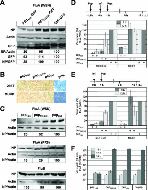
Peptide-specific inhibition of influenza A virus growth. (A) HEK293T cells were transfected with plasmids expressing the indicated GFP fusion proteins. After 24 h, cells were infected with influenza A virus (FluA) A/WSN/33 (WSN) at an MOI of 0.02. Western blot analysis was carried out 12 h p.i. with total cell extract to determine the levels of the viral NP, actin, and the GFP fusion proteins by using specific antibodies recognizing these proteins. Results from duplicate experiments are shown. The relative ratios of NP and actin signal intensities (NP/actin), the relative GFP signal intensities, and the relative ratios of NP and GFP signal intensities (NP/GFP) are shown. The ratio for cells expressing ctrl-GFP was set at 100. (B) HEK293T (293T) and MDCK cells were incubated with medium containing 10% fetal calf serum and 100 and 300 μM peptides, respectively, consisting of the transmembrane spanning region of TAT (aa 47 to 59) fused to aa 1 to 25 (pep1-25) or 715 to 740 (pep715-740) of PB1 or 25 aa of an unrelated viral sequence (pepCtrl). After 9 h (HEK293T) and 24 h (MDCK), cells were incubated with trypan blue solution (0.4%) for 5 min and washed two times with phosphate-buffered saline. Pos., positive control comprising Triton X-100-damaged cells. (C) HEK293T cells were incubated with medium containing 10% fetal calf serum and 100 μM peptides as indicated. After 1.5 h, cells were washed with phosphate-buffered saline and infected with influenza A virus strain A/WSN/33 for 4 h (upper panel) or A/PR8/34 for 5 h (middle panel) or influenza B virus (B/Lee/40) for 8 h (lower panel) in medium containing 2% fetal calf serum at an MOI of 0.2. Western blot analysis was performed using total cell extract and influenza A and B virus-specific anti-NP and anti-actin antibodies. Results from duplicate experiments are shown. The means of the relative ratios of NP and actin signal intensities are shown, and the ratios observed for cells incubated with pep-ctrl were set at 100. (D) MDCK cells were incubated with 300 μM peptides (Pep., −1.5 h) and infected (Inf., 0 h) with influenza A virus (A/WSN/33) as described in the legend to panel B at an MOI of 0.02 or 2. One hour p.i., cells were washed and further incubated in culture medium containing 2% fetal calf serum and 300 μM peptides (Pep., 1 h) as indicated. The viral titers in the cell culture supernatants were determined by plaque assays. The relative virus yields from pep-ctrl-treated cells observed at 9 and 12 h p.i., corresponding to 2 × 10⁴ and 3 × 10⁵ PFU/ml at an MOI of 0.02 and 5 × 10⁴ and 3 × 10⁵ PFU/ml at an MOI of 2, respectively, were set at 100%. Experiments were performed with triplicate assays. Error bars represent standard deviations of the mean values. +, present. (E) MDCK cells were infected with influenza A virus (A/WSN/33) at an MOI of 0.02 or 2 as described in the legend to panel D but without preincubation with peptides. The viral titers in the cell culture supernatants were determined by plaque assays. The relative virus yields from pep-ctrl-treated cells observed at 9 and 12 h p.i., corresponding to 1 × 10³ and 1 × 10⁴ PFU/ml at an MOI of 0.02 and 5 × 10⁴ and 3 × 10⁵ PFU/ml at an MOI of 2, respectively, were set at 100%. (F) MDCK cells were infected with influenza A virus (A/SC35M) at an MOI of 0.02 and treated with peptides as described in the legend to panel E. Stars indicate values from early time points at which titers could be detected in only one of three independent experiments.
References
-
- Crescenzo-Chaigne, B., N. Naffakh, and S. van der Werf. 1999. Comparative analysis of the ability of the polymerase complexes of influenza viruses type A, B and C to assemble into functional RNPs that allow expression and replication of heterotypic model RNA templates in vivo. Virology 265:342-353. - PubMed
-
- Dostmann, W. R., C. Nickl, S. Thiel, I. Tsigelny, R. Frank, and W. J. Tegge. 1999. Delineation of selective cyclic GMP-dependent protein kinase Ialpha substrate and inhibitor peptides based on combinatorial peptide libraries on paper. Pharmacol. Ther. 82:373-387. - PubMed
Publication types
MeSH terms
Substances
LinkOut - more resources
Full Text Sources
Other Literature Sources
Miscellaneous
