. 2007 Jul;81(14):7504-16.
doi: 10.1128/JVI.02690-06. Epub 2007 May 9.

Poliovirus induces Bax-dependent cell death mediated by c-Jun NH2-terminal kinase

Affiliations

Poliovirus induces Bax-dependent cell death mediated by c-Jun NH2-terminal kinase

Arnaud Autret et al. J Virol. 2007 Jul.

Abstract

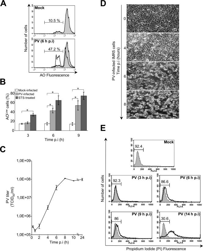
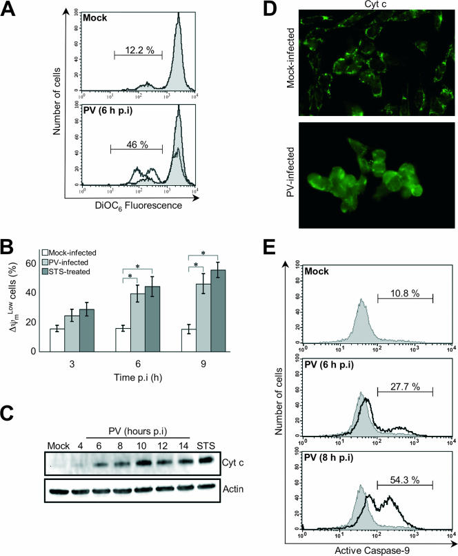
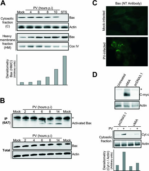
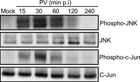
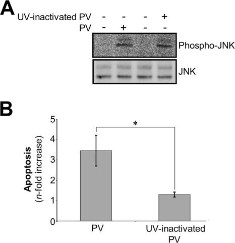
Poliovirus (PV) is the causal agent of paralytic poliomyelitis, a disease that involves the destruction of motor neurons associated with PV replication. In PV-infected mice, motor neurons die through an apoptotic process. However, mechanisms by which PV induces cell death in neuronal cells remain unclear. Here, we demonstrate that PV infection of neuronal IMR5 cells induces cytochrome c release from mitochondria and loss of mitochondrial transmembrane potential, both of which are evidence of mitochondrial outer membrane permeabilization. PV infection also activates Bax, a proapoptotic member of the Bcl-2 family; this activation involves its conformational change and its redistribution from the cytosol to mitochondria. Neutralization of Bax by vMIA protein expression prevents cytochrome c release, consistent with a contribution of PV-induced Bax activation to mitochondrial outer membrane permeabilization. Interestingly, we also found that c-Jun NH(2)-terminal kinase (JNK) is activated soon after PV infection and that the PV-cell receptor interaction alone is sufficient to induce JNK activation. Moreover, the pharmacological inhibition of JNK by SP600125 inhibits Bax activation and cytochrome c release. This is, to our knowledge, the first demonstration of JNK-mediated Bax-dependent apoptosis in PV-infected cells. Our findings contribute to our understanding of poliomyelitis pathogenesis at the cellular level.


Figures

FIG. 1.
FIG. 1.
PV-induced apoptosis in neuronal cells. (A) Representative flow cytometric histograms after AO nuclear dye staining of mock-infected and PV-infected (6 h p.i.) IMR5 cells. The profiles of mock-infected control cells (gray area) and PV-infected cells (blank area) are shown. The percentages of apoptotic cells corresponding to a reduced fluorescence intensity (AOLow) for each of the two experimental conditions are indicated. (B) Flow cytometric analysis of PV-induced apoptosis. Mock-infected, PV-infected, and STS-treated IMR5 cells were analyzed at the indicated times p.i. by flow cytometry after AO staining, and the percentages of AOLow cells are shown in white, light gray, and dark gray, respectively. The graph shows the mean percentages of apoptotic cells in three independent experiments. Error bars represent the standard errors of the means. *, P < 0.05 by Student's t test comparing PV-infected and STS-treated IMR5 cells to mock-infected IMR5 cells. (C) One-step growth curve of PV in IMR5 cells. Cells and supernatants were harvested at the indicated times p.i. and subjected to three cycles of freezing and thawing. Total virus yields were determined by TCID50 assay. Each point represents the mean virus titer for three independent experiments. Standard errors of the means are indicated. (D) Kinetics of cytopathic effect in PV-infected IMR5 cells. Cells were visualized by light microscopy at the indicated times p.i. Magnification, ×150. (E) Assessment of plasma membrane integrity. Flow cytometry histograms produced after staining of mock- and PV-infected IMR5 cells with PI at the indicated times. PI cannot enter cells with intact plasma membranes. When the plasma membranes are disrupted, cells become permeable to PI. The percentages of cells with intact membranes are indicated. Diagrams representative of two experiments are shown.
FIG. 2.
FIG. 2.
PV-induced mitochondrial damage in neuronal cells. (A) Representative flow cytometric histograms after DiOC6 staining of mock-infected (gray area) and PV-infected (6 h p.i.; white area) IMR5 cells. The percentages of apoptotic cells corresponding to a reduced fluorescence intensity (ΔψmLow) for each of the two experimental conditions are indicated. (B) Flow cytometric analysis of mitochondrial-permeability transition in PV-infected IMR5 cells. Mock-infected, PV-infected, and STS-treated IMR5 cells were analyzed at the indicated times p.i. by flow cytometry after DiOC6 staining, and the percentages of ΔψmLow cells are shown in white, light gray, and dark gray, respectively. The graph reports the mean percentages of ΔψmLow cells obtained from three independent experiments. Error bars represent the standard errors of the means. *, P < 0.05 by Student's t test comparing PV-infected and STS-treated IMR5 cells to mock-infected IMR5 cells. (C) Time course of cytochrome c redistribution in PV-infected IMR5 cells. At the indicated times p.i., cells were collected and subjected to subcellular fractionation. The cytochrome c (Cyt c) was detected in the cytosolic fraction by Western blotting analysis with an anti-cytochrome c antibody. Mock-infected IMR5 cells and cells treated with STS for 8 h were used as negative and positive controls, respectively. Levels of actin were used to control for protein loading. (D) Cytochrome c redistribution in PV-infected IMR5 cells. Mock-infected and PV-infected IMR5 (6 h p.i.) cells were stained by immunofluorescence with a specific monoclonal antibody against cytochrome c and a secondary, fluorescein-conjugated antibody. See the text for details. (E) Caspase-9 activation in PV-infected IMR5 cells. Caspase-9 activation was determined in mock- and PV-infected IMR5 cells (6 h and 8 h p.i.) by flow cytometry, using a fluorescein-labeled inhibitor (FAM-LEHD-fmk) that binds specifically to active caspase-9, as described in Materials and Methods. A histogram representative of two independent experiments is shown. The percentages of cells positive for activated caspase-9 are indicated.
FIG. 3.
FIG. 3.
PV-mediated cytochrome c release in neuronal cells is Bax dependent. (A) Translocation of the proapoptotic protein Bax to mitochondria in PV-infected IMR5 cells. At the indicated time p.i., equal amounts of cytosolic and mitochondrial proteins were assayed for Bax by Western blotting. Mock-infected and STS-treated (8 h posttreatment) IMR5 cells were used as negative and positive controls, respectively. Cox IV and actin were used as protein loading controls for heavy membrane and cytosolic fractions, respectively. Protein levels of heavy membrane and cytosolic fractions were determined by densitometry and plotted as ratios relative to the levels of Cox IV and actin, respectively. (B) Time course of PV-induced Bax conformational change in IMR5 cells. (Top) Mock-infected and PV-infected IMR5 cells were lysed in immunoprecipitation buffer. Conformationally active Bax protein was immunoprecipitated (IP) with anti-Bax 6A7 antibody and the precipitates were immunoblotted with anti-Bax antibody. The asterisk indicates immunoglobulin light chains. (Bottom) Whole-cell lysates that were not incubated with antibody were similarly tested for Bax by immunoblotting to check for equal amounts of Bax protein in samples prior to immunoprecipitation. Actin was used as protein loading controls. (C) PV-induced Bax activation. Mock-infected and PV-infected (6 h p.i.) IMR5 cells were analyzed by immunofluorescence with an antibody specific for the N terminus of Bax (NT antibody) to detect Bax conformational change. (D) Inhibition of cytochrome c release after vMIA expression in PV-infected IMR5 cells. (Top) Cells were transfected with plasmids encoding Myc-tagged vMIA, were transfected with empty vector (pcDNA3.1), or were left untreated. vMIA protein was detected in whole-cell lysates by Western blotting analysis with anti-Myc antibody. Actin was used as a protein loading control. The asterisk indicates a probable nonspecific protein band. (Bottom) Cytochrome c (Cyt c) release 24 h after transfection was analyzed in cytosolic fractions of mock-infected and PV-infected cells (8 h p.i.) by immunoblotting. Protein levels were determined by densitometry and plotted as ratios relative to the levels of actin.
FIG. 4.
FIG. 4.
PV-mediated MOMP in neuronal cells is Bid and Bim independent. (A) Time course of caspase-8 and Bid processing in PV-infected IMR5 cells. (Top) At the indicated times p.i., whole-cell extracts were subjected to immunoblot analysis with anti-caspase-8 (Pro-C8) and anti-Bid antibodies. Actin was used as a control for protein loading. (Bottom) Caspase-8 activation in PV-infected IMR5 cells. Caspase-8 activation was determined in mock- and PV-infected IMR5 cells (14 h p.i.) by flow cytometry using a fluorescein-labeled inhibitor (FAM-LETD-fmk) that binds specifically to active caspase-8, as described in Materials and Methods. Histograms representative of two independent experiments are shown. The percentages of cells positive for activated caspase-8 are indicated. (B) The broad-spectrum caspase inhibitor z-VAD-fmk does not inhibit cytochrome c release during PV infection. Mock- and PV-infected IMR5 cells were left untreated or treated with 100 μM z-VAD-fmk for 2 h before PV infection, and the inhibitor concentration was maintained during the adsorption period and throughout PV infection. Cytochrome c (Cyt c) was assayed in cytosolic extracts by Western blot analysis (14 h p.i.). Actin was used as a control for protein loading. (C) No inhibition of cytochrome c release after knockdown of Bim expression in PV-infected cells. (Top) IMR5 cells were transfected with Bim siRNA or nontargeted control siRNA. Bim protein was then assayed by immunoblotting with extracts from nontargeted control siRNA-transfected and Bim siRNA-transfected cells. Actin was used as a protein loading control. (Bottom) Cells were uninfected or were infected with PV 72 h after transfection, and cytochrome c (Cyt c) release was analyzed in cytosolic fractions by Western blotting 8 h p.i. Actin was used as a protein loading control. Protein levels were determined by densitometry and plotted as ratios relative to the levels of actin.
FIG. 5.
FIG. 5.
Early activation of JNK and subsequent c-Jun phosphorylation in PV-infected neuronal cells. Activation of each JNK and c-Jun was analyzed in whole-cell lysates at the indicated times p.i. by Western blotting using specific anti-phospho (Thr183/Tyr185)-JNK (p46 [JNK1] and p54 [JNK2/3]) and anti-phospho (Ser63)-c-Jun antibodies. Blots were subsequently stripped and reprobed with antibodies recognizing all JNK and c-Jun forms to confirm equal protein loading.
FIG. 6.
FIG. 6.
Bax translocation, cytochrome c release, and cell death following PV infection depend on JNK activation. (A) Inhibition of JNK activity during PV infection in IMR5 cells treated with SP600125 (25 μM). Cells were incubated with the JNK inhibitor for 2 h before PV infection, and the inhibitor concentration was maintained during the adsorption period and throughout PV infection. Levels of phospho (Thr183/Tyr185)-JNK and phospho (Ser63)-c-Jun in whole-cell lysates were determined at 30 min p.i. by Western blotting. Blots were subsequently stripped and reprobed with antibodies recognizing all JNK and c-Jun forms to confirm equal protein loading. (B) IMR5 cells were uninfected or were infected with PV in the presence or absence of SP600125 (25 μM). Whole-cell lysates (8 h p.i.) were subjected to Western blot analysis with anti-Bax and anti-cytochrome c (Cyt c) antibodies. Actin was used as a control for protein loading. (C) Inhibition of Bax translocation from the cytosol to mitochondria by the JNK inhibitor SP600125. IMR5 cells were uninfected or were infected with PV in the presence or the absence of SP600125 (25 μM). Eight hours p.i., the cytosolic and heavy membrane fractions were assayed for Bax by Western blotting. Actin and Cox IV were used as controls for protein loading of cytosolic and heavy membrane fractions, respectively. Protein levels of heavy membrane and cytosolic fractions were determined by densitometry and plotted as ratios relative to the levels of Cox IV and actin, respectively. (D) Cytochrome c release is reduced by JNK inhibitor. IMR5 cells were uninfected or were infected with PV in the presence or absence of SP600125 (25 μM). Cytosolic extract proteins were analyzed at 14 h p.i. by immunoblotting with anti-cytochrome c (Cyt c) antibody. Actin was used as a control for protein loading. Protein levels were determined by densitometry and plotted as ratios relative to the levels of actin. (E) Inhibition of PV-induced cytopathic effect by the JNK inhibitor SP600125. Cells were uninfected or were infected with PV in the presence or absence of SP600125 (25 μM) and visualized by light microscopy at the indicated times p.i. Magnification, ×350. (F) The JNK inhibitor SP600125 does not affect PV growth but affects PV release. IMR5 cells were infected with PV in the presence or absence of SP600125 (25 μM). Total virus yield (extracellular and intracellular) was determined by TCID50 assay at the indicated times after three cycles of freezing and thawing to release intracellular viruses. The titers of extracellular virus were determined from the supernatant of PV-infected cells at the indicated times after the removal of detached cells by centrifugation. Each point represents the mean virus titer for two independent experiments. Standard errors of the means are indicated. *, P < 0.05 by Student's t test comparing untreated IMR5 cells to treated IMR5 cells.
FIG. 7.
FIG. 7.
UV-inactivated PV induces early JNK activation but not cell death in IMR5 cells. (A) UV-inactivated PV induces early JNK activation in IMR5 cells. JNK activation was analyzed by Western blotting whole-cell lysates from cells infected with infectious or UV-inactivated PV (30 min p.i.) with specific anti-phospho (Thr183/Tyr185)-JNK antibody. Blots were subsequently stripped and reprobed with antibodies recognizing all forms of JNK to confirm equal protein loading. (B) UV-inactivated PV did not induce cell death in IMR5 cells. Mock-infected IMR5 cells and cells infected with infectious PV or UV-inactivated PV (8 h p.i.) were analyzed by flow cytometry after AO staining and the increase (n-fold) in apoptosis was calculated as the ratio of the percentage of apoptotic cells among PV-infected IMR5 cells to the percentage of apoptotic cells among mock-infected IMR5 cells. Data are means from two independent experiments. Error bars represent the standard errors of the means. *, P < 0.05 by Student's t test comparing untreated IMR5 cells to treated IMR5 cells.

Similar articles

Cited by

References

    1. Agol, V. I., G. A. Belov, K. Bienz, D. Egger, M. S. Kolesnikova, L. I. Romanova, L. V. Sladkova, and E. A. Tolskaya. 2000. Competing death programs in poliovirus-infected cells: commitment switch in the middle of the infectious cycle. J. Virol. 74:5534-5541. - PMC - PubMed
    1. Ammendolia, M. G., A. Tinari, A. Calcabrini, and F. Superti. 1999. Poliovirus infection induces apoptosis in CaCo-2 cells. J. Med. Virol. 59:122-129. - PubMed
    1. Ammendrup, A., A. Maillard, K. Nielsen, N. Aabenhus Andersen, P. Serup, O. Dragsbaek Madsen, T. Mandrup-Poulsen, and C. Bonny. 2000. The c-Jun amino-terminal kinase pathway is preferentially activated by interleukin-1 and controls apoptosis in differentiating pancreatic beta-cells. Diabetes 49:1468-1476. - PubMed
    1. Aoki, H., P. M. Kang, J. Hampe, K. Yoshimura, T. Noma, M. Matsuzaki, and S. Izumo. 2002. Direct activation of mitochondrial apoptosis machinery by c-Jun N-terminal kinase in adult cardiac myocytes. J. Biol. Chem. 277:10244-10250. - PubMed
    1. Arnoult, D., L. M. Bartle, A. Skaletskaya, D. Poncet, N. Zamzami, P. U. Park, J. Sharpe, R. J. Youle, and V. S. Goldmacher. 2004. Cytomegalovirus cell death suppressor vMIA blocks Bax- but not Bak-mediated apoptosis by binding and sequestering Bax at mitochondria. Proc. Natl. Acad. Sci. USA 101:7988-7993. - PMC - PubMed

Publication types

Substances