. 2007 Jul;81(14):7371-9.
doi: 10.1128/JVI.00513-07. Epub 2007 May 9.

CD163 expression confers susceptibility to porcine reproductive and respiratory syndrome viruses

Affiliations

CD163 expression confers susceptibility to porcine reproductive and respiratory syndrome viruses

Jay G Calvert et al. J Virol. 2007 Jul.

Abstract

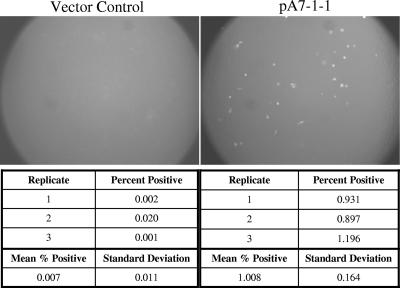
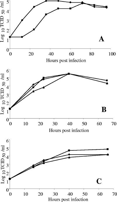
Direct functional screening of a cDNA expression library derived from primary porcine alveolar macrophages (PAM) revealed that CD163 is capable of conferring a porcine reproductive and respiratory syndrome virus (PRRSV)-permissive phenotype when introduced into nonpermissive cells. Transient-transfection experiments showed that full-length CD163 cDNAs from PAM, human U937 cells (histiocytic lymphoma), African green monkey kidney cells (MARC-145 and Vero), primary mouse peritoneal macrophages, and canine DH82 (histocytosis) cells encode functional virus receptors. In contrast, CD163 splice variants without the C-terminal transmembrane anchor domain do not provide PRRSV receptor function. We established several stable cell lines expressing CD163 cDNAs from pig, human, and monkey, using porcine kidney (PK 032495), feline kidney (NLFK), or baby hamster kidney (BHK-21) as the parental cell lines. These stable cell lines were susceptible to PRRSV infection and yielded high titers of progeny virus. Cell lines were phenotypically stable over 80 cell passages, and PRRSV could be serially passed at least 60 times, yielding in excess of 10(5) 50% tissue culture infective doses/ml.


Figures

FIG. 1.
FIG. 1.
Constructs used for generating cell lines FK-D4 (feline kidney cells expressing simian CD163) and PK-9 (porcine kidney cells expressing porcine CD163). (A) The RSV promoter (pRSV) and neomycin resistance cassette were isolated from the pRSV-Script vector. Separately, the MARC-CD163v2 gene with a NotI 5′ restriction endonuclease recognition site was removed from pCDNA3.1D. The in vitro-ligated materials were separated on a preparative agarose gel. Linear DNA containing the CD163 gene under control of the RSV promoter was excised from the gel. The purified DNA was used for transfection of NLFK cells. (B) A circular pCMV-Script plasmid containing susCD163v1 and a neomycin resistance cassette was used for transfection of PK032495 cells. SV40pA, simian virus 40 polyadenylation signal; pSV40, simian virus 40 promoter; pCMV, human CMV immediate-early promoter.
FIG. 2.
FIG. 2.
PAM library clone A7-1-1 contains a putative PRRSV receptor. BHK-21 cells were transiently transfected (four replicates) with either the original PAM library-derived CD163-containing clone (A7-1-1) or a vector control plasmid (pPAMB) containing a small irrelevant PAM library insert. Following overnight incubation, the transfection mixture was removed and replaced with fresh medium. After several hours at 37°C, this medium was removed and cells were infected with PRRSV P129-GFP. At 1 day postinfection, three of the replicate wells were harvested for flow cytometry (cell counts, below) and the remaining well was analyzed by fluorescence microscopy (photomicrographs, above).
FIG. 3.
FIG. 3.
Structural domain organization of CD163 proteins. The deletion of two SRCR domains in variant susCD163v1 (middle) is compared to full-length susCD163v2 (top). A variant lacking the transmembrane domain is represented by VeroCD163 (TM −) (bottom).
FIG. 4.
FIG. 4.
Growth of PRRSV on stably transfected cells expressing CD163. Replicate wells of PK-9 cells were infected with P129/p16 (passaged 16 times in PK-9 cells). Cells were fixed daily with 80% acetone and stained with FITC-conjugated MAb SDOW-17. The PRRSV N protein was visualized by fluorescence microscopy. Development of infection started with a few infected cells on day 1 (A), followed by enlargement of foci on day 2 (B). By day 3 postinfection, approximately 80% of the cells were infected.
FIG. 5.
FIG. 5.
Time courses of PRRSV replication on PK-9 and FK-D4 cells. (A) Growth kinetics were determined for PRRSV isolate P129 (passage 19 on PK-9 cells) using a multiplicity of 0.1 (⧫) and a multiplicity of 0.001 (▪) and titrated on PK-9 cells. After adsorption, inocula were removed and cells were washed with fresh medium. Culture fluids were harvested at 12-h intervals and virus titers were determined. (B) Growth curves for PRRSV P129/p38 (passage 38 on FK-D4 cells) were determined on FK-D4 cells at three passage levels to show stability of the PRRSV-permissive phenotype. Cells were evaluated at passage 19 (⧫), passage 29 (▪), and passage 46 (▴). Culture fluids were titrated on PAM cells. (C) Growth curves for P3412/p17 (passage 17 on FK-D4 cells) were evaluated on FK-D4 cells at passage 19 (⧫), passage 29 (▪), and passage 46 (▴). Inocula were removed after adsorption, and cells were washed with fresh medium. Culture fluids were titrated on PAM cells.
FIG. 6.
FIG. 6.
Blocking PRRSV infection with anti-human CD163 antibody. Infected cells were trypsinized, washed with PBS, and analyzed by flow cytometry, with at least 100,000 cells analyzed per sample. (A) Recombinant FK-A6 cells stably expressing humCD163v2 were incubated with either goat anti-human CD163-specific antibody (▴) or normal goat IgG (▪) and infected with P129-GFP virus. At 24 h postinfection, the percentage of GFP-expressing cells was determined by flow cytometry. (B) MARC-145 cells were incubated with either goat anti-human CD163-specific antibody (▴) or normal goat IgG (▪) and infected with P129-GFP virus. At 24 h postinfection, the percentage of GFP-expressing cells was determined by flow cytometry. Error bars represent the standard deviation of three replicate counts.
FIG. 7.
FIG. 7.
Western blot analysis of CD163 proteins. Lysates of MARC-145, FK-A6.A2, FK-D4, or parental NLFK were separated under reducing conditions. Proteins were transferred to a polyvinylidene difluoride membrane and incubated with goat anti-human CD163 polyclonal antibody followed by rabbit anti-goat antibody labeled with alkaline phosphatase. Membranes were developed in a Western Blue alkaline phosphatase substrate. Arrows indicate the CD163 protein at approximately 130 kDa. MW, prestained molecular mass markers (Invitrogen).
FIG. 8.
FIG. 8.
Detection of sialoadhesin mRNA. A primer pair located at the 3′ end of the sialoadhesin gene was used in RT-PCRs to amplify sialoadhesin mRNA from PAM, PK-15, and PK032495 cells. RT-PCR products were separated on a preformed 0.8% E-gel (Invitrogen) and visualized under the UV light. A 469-bp RT-PCR fragment was amplified from PAM cells (arrow) but not from either PK cell line. M, Ready Load 1-kb ladder (Invitrogen).

Similar articles

Cited by

References

    1. Cavanagh, D. 1997. Nidovirales: a new order comprising Coronaviridae and Arteriviridae. Arch. Virol. 142:629-633. - PubMed
    1. Delputte, P. L., S. Costers, and H. J. Nauwynck. 2005. Analysis of porcine reproductive and respiratory syndrome virus attachment and internalization: distinctive roles for heparin sulfate and sialoadhesin. J. Gen. Virol. 86:1441-1445. - PubMed
    1. Delputte, P. L., N. Vanderheijden, H. J. Nauwynck, and M. B. Pensaert. 2002. Involvement of the matrix protein in attachment of porcine reproductive and respiratory syndrome virus to a heparin like receptor on porcine alveolar macrophages. J. Virol. 76:4312-4320. - PMC - PubMed
    1. Duan, X., H. J. Nauwynck, and M. B. Pensaert. 1997. Effects of origin and state of differentiation and activation of monocytes/macrophages on their susceptibility to porcine reproductive and respiratory syndrome virus. Arch. Virol. 142:2483-2497. - PMC - PubMed
    1. Duan, X., H. J. Nauwynck, and M. B. Pensaert. 1997. Virus quantification and identification of cellular targets in the lungs and lymphoid tissues of pigs at different time intervals after inoculation with porcine reproductive and respiratory syndrome virus. Vet. Microbiol. 56:9-19. - PubMed

Publication types

MeSH terms