

3D digital subtraction angiography of intracranial aneurysms: comparison of flat panel detector with conventional image intensifier TV system using a vascular phantom
- PMID: 17494653
- PMCID: PMC8134332
3D digital subtraction angiography of intracranial aneurysms: comparison of flat panel detector with conventional image intensifier TV system using a vascular phantom
Abstract
Background and purpose: Compared with the image intensifier (I.I.)-TV system, the flat panel detector (FPD) system of direct conversion type has several theoretic advantages, such as higher spatial resolution, wide dynamic range, and no image distortion. The purpose of this study was to compare the image quality of 3D digital subtraction angiography (DSA) in the FPD and conventional I.I.-TV systems using a vascular phantom.
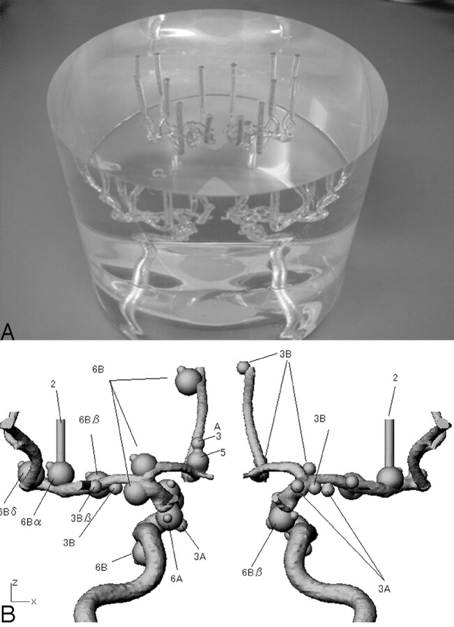
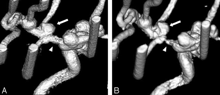
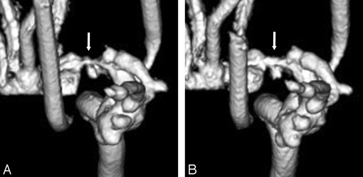
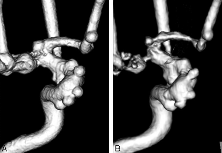
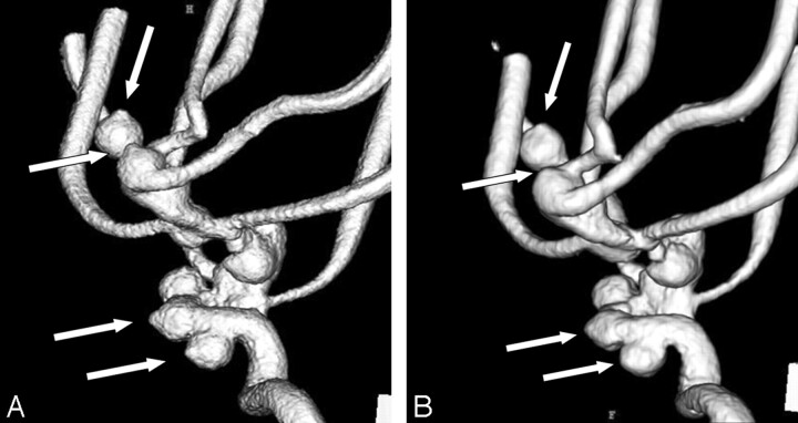
Materials and methods: An anthropomorphic vascular phantom was designed to simulate the various intracranial aneurysms with aneurysmal bleb. The tubes of this vascular phantom were filled with 2 concentrations of contrast material (300 and 150 mg I/mL), and we obtained 3D DSA using the FPD and I.I.-TV systems. First, 2 blinded radiologists compared the volume-rendering images for 3D DSA on the FPD and I.I.-TV systems, looking for pseudostenosis artifacts. Then, 2 other radiologists independently evaluated both systems for the depiction of the simulated aneurysm and aneurysmal bleb using a 5-point scale.
Results: For the degree of the pseudostenosis artifacts at the M1 segment of the middle cerebral artery at 300 mg I/mL, 3D DSA with FPD system showed mild stenoses, whereas severe stenoses were observed at 3D DSA with I.I.-TV system. At both concentrations, the FPD system was significantly superior to I.I.-TV system regarding the depiction of aneurysm and aneurysmal bleb.
Conclusion: Compared with the I.I.-TV system, the FPD system could create high-resolution 3D DSA combined with a reduction of the pseudostenosis artifacts.
Figures
References
-
- Anxionnat R, Bracard S, Ducrocq X, et al. Intracranial aneurysms: clinical value of 3D digital subtraction angiography in the therapeutic decision and endovascular treatment. Radiology 2001;218:799–808 - PubMed
-
- Tanoue S, Kiyosue H, Kenai H, et al. Three-dimensional reconstructed images after rotational angiography in the evaluation of intracranial aneurysm: surgical correlation. Neurosurgery 2000;47:866–71 - PubMed
-
- Anxionnat R, Bracard S, Macho J, et al. 3D angiography: clinical interest—first applications in interventional neuroradiology. J Neuroradiol 1998;25:251–62 - PubMed
-
- Hirai T, Korogi Y, Ono K, et al. Pseudostenosis phenomenon at volume-rendered three-dimensional digital angiography of intracranial arteries: frequency, location, and effect on image evaluation. Radiology 2004;232:882–87 - PubMed
Publication types
MeSH terms
LinkOut - more resources
Full Text Sources
Medical