

Case Reports
Artery of Percheron thrombolysis
Affiliations
- PMID: 17494659
- PMCID: PMC8134353
Item in Clipboard
Case Reports
Artery of Percheron thrombolysis
AJNR Am J Neuroradiol.
2007 May.
Abstract
A patient with acute top of the basilar syndrome clinically was found to have only a small basilar artery filling defect but complete occlusion of the artery of Percheron. Intra-arterial thrombolysis resulted in favorable neurologic outcome. To our knowledge, this is the only case of angiographically proved and treated artery of Percheron occlusion. The value of this report is that reperfusion of ischemic areas was only achieved when persistent investigation disclosed artery of Percheron occlusion.
Figures
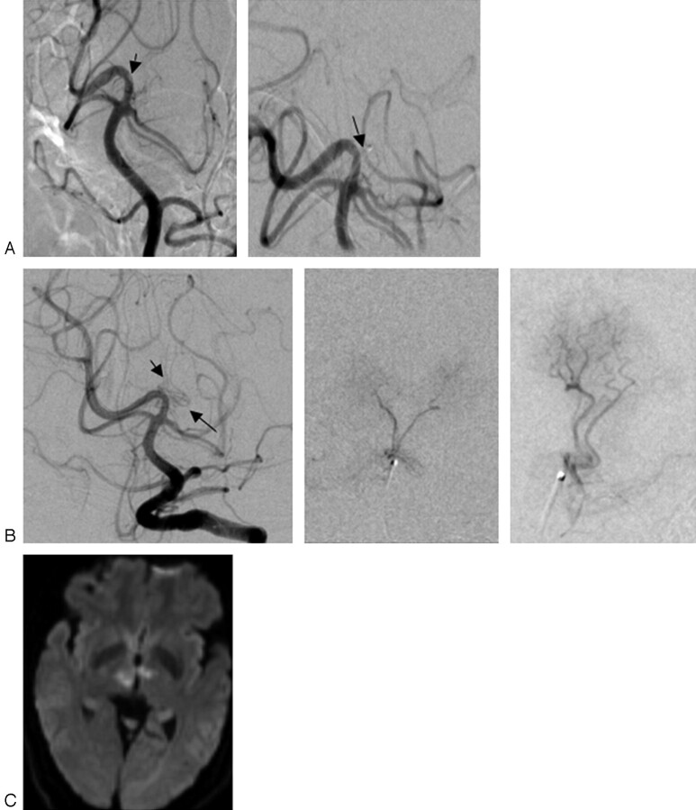
A, Prethrombolysis. Left: The left vertebral artery angiogram shows a small filling defect (black arrow). The left P1 segment of the posterior cerebral artery is aplastic in this patient; thus, the defect could be either the basilar artery tip or the proximal-most right P1 segment. Right: Selective angiogram shows the microcatheter extending beyond the right P1 segment into the proximal lumen of the occluded artery of Percheron. Arrow indicates catheter tip. B, Post-thrombolysis. Left: The left vertebral artery angiogram shows normal visualization of the full length of the artery of Percheron (arrows) as does microcatheter selective angiograms of the artery of Percheron at center and right. The proximal occlusion of the artery of Percheron seen in Fig 1A is no longer present. C, DWI MR image obtained 24 hours after intervention shows minimal final ischemic injury.
References
-
- The Editorial Committee for the Guarantors of Brain. Aids to the Examination of the Peripheral Nervous System. London: Billiere Tindall,1986. .
-
- Percheron G. Arteries of the human thalamus. I. Artery and polar thalamic territory of the posterior communicating artery [in French]. Rev Neurol (Paris) 1976;132:297–307 - PubMed
-
- Kumral E, Evyapan D, Balkir K, et al. Bilateral thalamic infarction: clinical, etiological and MRI correlates. Acta Neurol Scand 2001;103:35–42 - PubMed
-
- Caplan LR. “Top of the basilar” syndrome. Neurology 1980;30:72–79 - PubMed
-
- Lepore FE, Gulli V, Miller DC. Neuro-ophthalmological findings with neuropathological correlation in bilateral thalamic-mesencephalic infarction. J Clin Neuroophthalmol 1985;5:224–28 - PubMed
Publication types
MeSH terms
Substances
LinkOut - more resources
Full Text Sources