Clinical Trial
. 2007 May;28(5):877-84.

MR angiography of dural arteriovenous fistulas: diagnosis and follow-up after treatment using a time-resolved 3D contrast-enhanced technique

Affiliations
Clinical Trial

MR angiography of dural arteriovenous fistulas: diagnosis and follow-up after treatment using a time-resolved 3D contrast-enhanced technique

S Meckel et al. AJNR Am J Neuroradiol. 2007 May.

Abstract

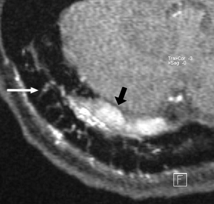
Background and purpose: Digital subtraction angiography (DSA) is the method of reference for imaging of dural arteriovenous fistula (DAVF). The goal of this study was to analyze the value of different MR images including 3D contrast-enhanced MR angiography (MRA) with a high temporal resolution in diagnostic and follow-up imaging of DAVFs.

Materials and methods: A total of 18 MR/MRA examinations from 14 patients with untreated (n=9) and/or treated (n=9) DAVFs were evaluated. Two observers assessed all MR and MRA investigations for signs indicating the presence of a DAVF, for fistula characteristics such as fistula grading, location of fistulous point, and fistula obliteration after treatment. All results were compared with DSA findings.

Results: On time-resolved 3D contrast-enhanced (TR 3D) MRA, the side and presence of all patent fistulas (n=13) were correctly indicated, and no false-positive findings were observed in occluded DAVFs (n=5). Grading of fistulas with this imaging technique was correct in 77% and 85% of patent fistulas for both readers, respectively. On T2-weighted images, signs indicative of a DAVF were encountered only in fistulas with cortical venous reflux (56%), whereas on 3D time-of-flight (TOF) MRA, most fistulas (88%) were correctly detected. In complete fistula occlusion, false-positive findings were encountered on both T2-weighted images and on TOF MRA images.

Conclusion: In this study, TR 3D MRA proved reliable in detecting DAVFs and suitable for follow-up imaging. The technique allowed--within limitations--to grade DAVFs. Although 3D TOF MRA can depict signs of DAVFs, its value for follow-up imaging is limited.


Figures

Fig 1.
Fig 1.
Targeted sagittal MIP reconstructed image of a TR MRA dataset obtained during early arterial phase (A) shows minimal venous filling of right sigmoid sinus (black arrow) in a patient with a low flow DAVF (type 1). The corresponding lateral DSA image with a right common carotid artery injection (B) demonstrates the fistula with mild early filling of right sigmoid sinus (black arrow) that is surrounded by a network of small feeding artery branches from the right occipital artery and middle meningeal artery (white arrowheads). This fistula was missed on 3D TOF MRA images.
Fig 2.
Fig 2.
Preinterventional targeted MIP reconstructed sagittal image from an arterial phase of TR 3D MRA (A) shows a DAVF at the transverse/sigmoid sinus (short arrow) with strong early filling of the sinuses and feeding arterial branches from a prominent right occipital artery (long arrow). Small reflux into the proximal part of the vein of Labbé (arrowhead) is noted. The targeted MIP reconstructed image obtained from follow-up TR 3D MRA examination (B) after transvenous occlusion of the right transverse sinus clearly shows a residual fistula at the sigmoid sinus (short arrow) with downstaging of venous hypertension to anterograde sinus flow (type 1).
Fig 3.
Fig 3.
Sagittal targeted MIP reconstructed image obtained from an early arterial phase of TR 3D MRA shows a DAVF with strong and early filling of the torcular of Herophili, of the right transverse/sigmoid sinus (long arrow) and intense venous reflux into an enlarged superior vermian vein (short arrow) that follows the course of the tentorium in the midline. A large network of feeding branches from bilateral occipital arteries is observed (arrowheads).
Fig 4.
Fig 4.
TR 3D MRA is obtained before and after surgical exclusion of a high-grade DAVF (type 4) on a left medial tentorial sinus. The complete arteriovenous series of sagittal MIP-reconstructed images (A) shows early enhancement of dilated veins with a large venous varix (black arrow) on the left tentorium at early arterial inflow (top left). Consecutive filling of the straight sinus and the transverse sinuses from the fistula is noted at later arterial phase (top right, black arrowheads). Regular temporal enhancement of the superior sagittal sinus (white arrowheads) can be appreciated during venous phases (lower panel). Note that fistula occlusion is definitely demonstrated by the absence of early filling of tentorial veins and varix on the follow-up study after surgery (B). On 3D TOF MRA, however, the venous varix (white arrow) is still depicted with a high signal intensity imitating fistula flow (C).
Fig 5.
Fig 5.
In a patient with a DAVF involving the right transverse sinus, transvenous coil occlusion of the distal part of the transverse sinus and the sigmoid sinus has been performed. A residual type 2a fistula at proximal part of the right transverse sinus with arterial feeders from the right occipital artery is demonstrated at follow-up imaging. The magnified axial source image of 3D TOF MRA shows multiple hyperintense thin transosseous vessels (white arrow) in the vicinity of the arterialized right transverse sinus (black arrow).

References

    1. Cognard C, Gobin YP, Pierot L, et al. Cerebral dural arteriovenous fistulas: clinical and angiographic correlation with a revised classification of venous drainage. Radiology 1995;194:671–80 - PubMed
    1. Davies MA, TerBrugge K, Willinsky R, et al. The validity of classification for the clinical presentation of intracranial dural arteriovenous fistulas. J Neurosurg 1996;85:830–37 - PubMed
    1. Borden JA, Wu JK, Shucart W. A proposed classification for spinal and cranial dural arteriovenous fistulous malformations and implications for treatment. J Neurosurg 1995;82:166–79 - PubMed
    1. Klisch J, Huppertz HJ, Spetzger U, et al. Transvenous treatment of carotid cavernous and dural arteriovenous fistulae: results for 31 patients and review of the literature. Neurosurgery 2003;53:836–56 - PubMed
    1. Goto K, Sidipratomo P, Ogata N, et al. Combining endovascular and neurosurgical treatments of high-risk dural arteriovenous fistulas in the lateral sinus and the confluence of sinuses. J Neurosurg 1999;90:289–99 - PubMed

LinkOut - more resources