

Cerebral proton magnetic resonance spectroscopy in children with diabetic ketoacidosis
- PMID: 17494665
- PMCID: PMC8134352
Cerebral proton magnetic resonance spectroscopy in children with diabetic ketoacidosis
Abstract
Background and purpose: Subclinical cerebral edema occurs in many, if not most, children with diabetic ketoacidosis (DKA) and may be an indicator of subtle brain injury. Brain ratios of N-acetylaspartate (NAA) to creatine (Cr), measured by proton MR spectroscopy, decrease with neuronal injury or dysfunction. We hypothesized that brain NAA/Cr ratios may be decreased in children in DKA, indicating subtle neuronal injury.
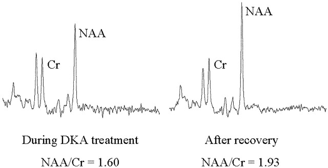
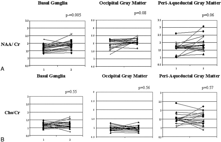
Materials and methods: Twenty-nine children with DKA underwent cerebral proton MR spectroscopy during DKA treatment (2-12 hours after initiating therapy) and after recovery from the episode (72 hours or more after the initiation of therapy). We measured peak heights of NAA, Cr, and choline (Cho) in 3 locations within the brain: the occipital gray matter, the basal ganglia, and periaqueductal gray matter. These regions were identified in previous studies as areas at greater risk for neurologic injury in DKA-related cerebral edema. We calculated the ratios of NAA/Cr and Cho/Cr and compared these ratios during the acute illness and recovery periods.
Results: In the basal ganglia, the ratio of NAA/Cr was significantly lower during DKA treatment compared with that after recovery (1.68 +/- 0.24 versus 1.86 +/- 0.28, P<.005). There was a trend toward lower NAA/Cr ratios during DKA treatment in the periaqueductal gray matter (1.66 +/- 0.38 versus 1.91 +/- 0.50, P=.06) and the occipital gray matter (1.97 +/- 0.28 versus 2.13 +/- 0.18, P=.08). In contrast, there were no significant changes in Cho/Cr ratios in any region.
Conclusions: NAA/Cr ratios are decreased in children during DKA and improve after recovery. This finding suggests that during DKA neuronal function or viability or both are compromised and improve after treatment and recovery.
Figures
References
-
- Glaser N, Barnett P, McCaslin I, et al. Risk factors for cerebral edema in children with diabetic ketoacidosis. N Engl J Med 2001;344:264–69 - PubMed
-
- Rosenbloom A. Intracerebral crises during treatment of diabetic ketoacidosis. Diabetes Care 1990;13:22–33 - PubMed
-
- Krane E, Rockoff M, Wallman J, et al. Subclinical brain swelling in children during treatment of diabetic ketoacidosis. N Engl J Med 1985;312:1147–51 - PubMed
Publication types
MeSH terms
Substances
LinkOut - more resources
Full Text Sources