

Diagnostic value of multidetector-row CT angiography in the evaluation of thrombosis of the cerebral venous sinuses
- PMID: 17494676
- PMCID: PMC8134361
Diagnostic value of multidetector-row CT angiography in the evaluation of thrombosis of the cerebral venous sinuses
Abstract
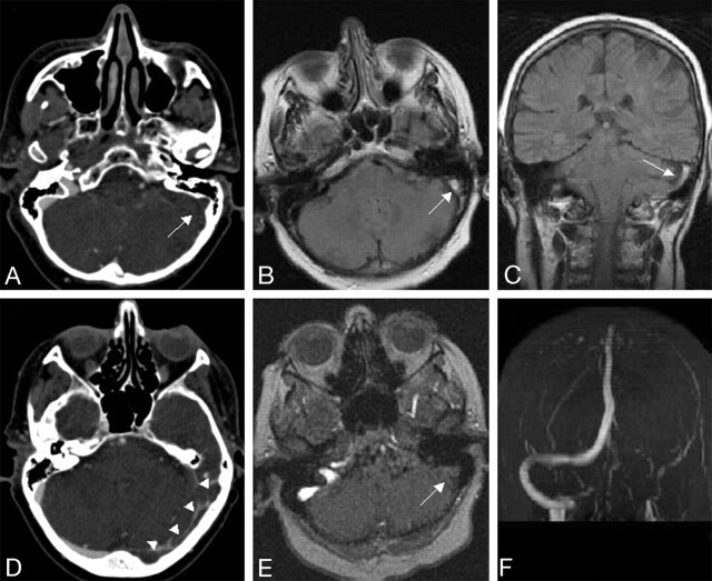
Background and purpose: The diagnosis of cerebral venous and sinus thrombosis (CVST) as a rare but important cause of stroke is challenging. We aimed to investigate the diagnostic value of multidetector-row CT angiography (MDCTA) as a fast and cost-effective imaging tool in diagnosing CVST.
Materials and methods: Nineteen patients who presented with clinical symptoms of a possible CVST were included. All patients had received both MDCTA and MR imaging with venous MR-angiography. Three blinded readers were asked to identify the cerebral sinuses and veins in MDCTA and to evaluate the presence of CVST in MDCTA. Consensus reading with interpretation of the MR imaging served to establish the definite diagnosis.
Results: The consensus reading revealed CVST in 10 of the 19 patients. With MDCTA, the venous sinuses could be identified in 99.2% and the cerebral veins in 87.6% of cases. The sensitivity and specificity of MDCTA for the diagnosis of CVST were 100%.
Conclusion: Our study demonstrates that MDCTA provides excellent sensitivity and specificity for the diagnosis of CVST. Further studies are needed to evaluate the diagnostic potential of MDCTA in specific subsets of the general entity of CVST such as cortical venous thrombosis, thrombosis of the cavernous sinus, and thrombosis of the internal cerebral veins.
Figures


Similar articles
-
Noncontrast CT in deep cerebral venous thrombosis and sinus thrombosis: comparison of its diagnostic value for both entities.AJNR Am J Neuroradiol. 2009 Apr;30(4):728-35. doi: 10.3174/ajnr.A1451. Epub 2009 Feb 12. AJNR Am J Neuroradiol. 2009. PMID: 19213820 Free PMC article.
-
Diagnostic role of 64-slice multidetector row CT scan and CT venogram in cases of cerebral venous thrombosis.Emerg Radiol. 2008 Sep;15(5):325-33. doi: 10.1007/s10140-008-0723-4. Epub 2008 Apr 24. Emerg Radiol. 2008. PMID: 18437434
-
[CT angiography of cerebral venous circulation: anatomical visualization and diagnostic pitfalls in interpretation].Rofo. 1997 Dec;167(6):612-8. doi: 10.1055/s-2007-1015591. Rofo. 1997. PMID: 9465957 German.
-
Complicated cerebral venous sinus thrombosis with intracranial hemorrhage and mastoiditis.Vasc Endovascular Surg. 2012 Oct;46(7):585-90. doi: 10.1177/1538574412457473. Epub 2012 Aug 22. Vasc Endovascular Surg. 2012. PMID: 22918938 Review.
-
Cerebral venous and dural sinus thrombosis* : state-of-the-art imaging.Clin Neuroradiol. 2010 Mar;20(1):25-37. doi: 10.1007/s00062-010-9035-7. Epub 2010 Feb 28. Clin Neuroradiol. 2010. PMID: 20229204 Review.
Cited by
-
Early Detection and Quantification of Cerebral Venous Thrombosis by Magnetic Resonance Black-Blood Thrombus Imaging.Stroke. 2016 Feb;47(2):404-9. doi: 10.1161/STROKEAHA.115.011369. Epub 2015 Dec 15. Stroke. 2016. PMID: 26670082 Free PMC article.
-
A Case of Cerebral Venous Sinus Thrombosis Presenting During Relapse of Ulcerative Colitis.Am J Case Rep. 2019 Mar 31;20:419-422. doi: 10.12659/AJCR.913429. Am J Case Rep. 2019. PMID: 30928992 Free PMC article.
-
Diagnostic accuracy and yield of multidetector CT angiography in the evaluation of spontaneous intraparenchymal cerebral hemorrhage.AJNR Am J Neuroradiol. 2009 Jun;30(6):1213-21. doi: 10.3174/ajnr.A1546. Epub 2009 Apr 2. AJNR Am J Neuroradiol. 2009. PMID: 19342546 Free PMC article.
-
Acute treatment of cerebral venous and dural sinus thrombosis.Curr Treat Options Neurol. 2008 Mar;10(2):126-37. doi: 10.1007/s11940-008-0014-0. Curr Treat Options Neurol. 2008. PMID: 18334135
-
Early imaging characteristics of 62 cases of cerebral venous sinus thrombosis.Exp Ther Med. 2013 Jan;5(1):233-236. doi: 10.3892/etm.2012.796. Epub 2012 Nov 2. Exp Ther Med. 2013. PMID: 23251274 Free PMC article.
References
-
- Bogousslavasky J, Pierre P. Ischaemic stroke in patients under age 45. Neurol Clin 1992;10:113–24 - PubMed
-
- Stam J. Thrombosis of the cerebral veins and sinuses. N Engl J Med 2005;352:1791–98 - PubMed
-
- Tehindrazanarivelo AD, Bousser MG. Idiopathic intracranial hypertension and cerebral dural sinus thrombosis. Am J Med 1994;97:200–01 - PubMed
-
- Leker RR, Steiner I. Features of dural sinus thrombosis simulating pseudotumour cerebri. Eur J Neurol 1999;6:601–04 - PubMed
-
- Renowden S. Cerebral venous sinus thrombosis. Eur Radiol 2004;14:215–26 - PubMed
MeSH terms
LinkOut - more resources
Full Text Sources
Medical