. 2007 May 9;27(19):5043-52.
doi: 10.1523/JNEUROSCI.5187-06.2007.

Rhythmic episodes of subthreshold membrane potential oscillations in the rat inferior olive nuclei in vivo

Affiliations

Rhythmic episodes of subthreshold membrane potential oscillations in the rat inferior olive nuclei in vivo

Edith Chorev et al. J Neurosci. .

Abstract

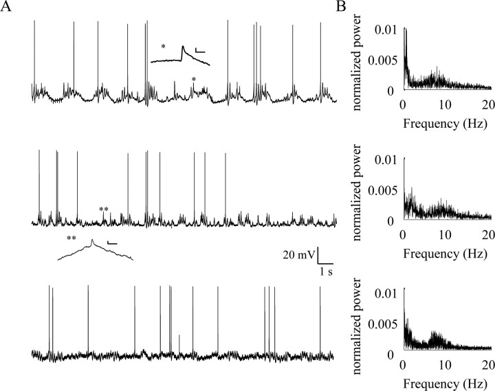
In vitro studies of inferior olive neurons demonstrate that they are intrinsically active, generating periodic spatiotemporal patterns. These self-generated patterns of activity extend the role of olivary neurons beyond that of a deliverer of teaching or error signals. However, autorhythmicity or patterned activity of complex spikes in the cerebellar cortex was observed in only a few studies. This discrepancy between the self-generated rhythmicity in the inferior olive observed in vitro and the sporadic reports on rhythmicity of complex spikes can be reconciled by recording intracellularly from inferior olive neurons in situ. To this end, we recorded intracellularly from olivary neurons of anesthetized rats. We demonstrate that, in vivo, olivary neurons show both slow and fast rhythmic processes. The slow process (0.2-2 Hz) is expressed as rhythmic transitions from quiescent periods to periods of fast rhythm, manifested as subthreshold oscillations of 6-12 Hz. Spikes, if they occur, are locked to the depolarized phase of these subthreshold oscillations and, therefore, hold and transfer rhythmic information. The transient nature of these oscillatory epochs accounts for the difficulties to uncover them by prolonged recordings of complex spikes activity in the cerebellar cortex.


Figures

Figure 1.
Figure 1.
Fluorescent labeling of the electrode tract and recording site. After olivary recording, we inserted an electrode filled with a fluorescent marker (RH414; Invitrogen, Eugene, OR) to the rostralmost coordinates we use for olivary recordings. The animal was then perfused with PBS and then with PFA; 100 μm slices were prepared and mounted on microscope slides. The labeling was detected using a fluorescence microscope. The marked areas are the site of penetration (the obex), the tract of the electrode and the injection site, which is at the rostralmost side of the inferior olive. Notice that the entire olive is marked by fluorescence. IO, Inferior olive; D, dorsal; V, ventral; C, caudal; R, rostral.
Figure 2.
Figure 2.
The rarity of 10 Hz rhythmicity in CS activity recorded from Purkinje cells. A, B, Two examples of CS activity recorded from Purkinje cells. Each example contains the raster plot (1), the CS and simple spikes wave forms (red and black traces; 2), autocorrelograms of the CS at slow (3) and fast (4) time scales, and the histogram of the ISI (5). Note that in both examples, a peak in the ISI histogram is evident (110 and 120 ms in A and B, respectively), but only in B a corresponding fast rhythm was detected in the autocorrelogram. A slow rhythm is also apparent only in B. CC, Correlation coefficient.
Figure 3.
Figure 3.
Two types of action potentials recorded from olivary neurons in vivo. A, Four superimposed high-threshold calcium spikes of various durations recorded from the inferior olivary neuron. Arrowheads denote wavelets that are likely to represent antidromic invasions of axonal spikes. B, Superposition of all spontaneous spikes from one experiment (82 spikes). The rising phase of all action potentials is shown in the inset. The arrow indicates the prepotentials that are presumably a low-threshold calcium component of the action potentials. Note that all of the action potentials are of the high-threshold type. C, Low-threshold action potential elicited by a square current pulse of 500 pA superimposed on a direct current hyperpolarization of 20 mV. D, The average ISI histogram from 32 neurons showing a peak at 95 ms (inset shows an enlargement of the area surrounding the peak of the histogram).
Figure 4.
Figure 4.
Spectral analysis of olivary neurons reveals two regions of increased power. A, Three examples of 20 s recordings from olivary neurons at their resting potential (−68, −60,−63 mV, top to bottom, respectively). B, The corresponding power spectra of the three examples shown in A. Two regions of increased power are seen in all three cases; the first region is between 0.2–2 Hz and the second between 5 and 13 Hz. Insets in A show an enlarged display of spikelet-like events (asterisks). Calibration: 2 mV, 5 ms. Note that the sharp peak of the slow rhythm in the first example is at 0.5 Hz. In this case, the animal was respirated at 1.6 Hz.
Figure 5.
Figure 5.
Suprathreshold activity shows two rhythmic processes. A, Twenty second recordings from three olivary neurons recorded in absence of injected current. B, The corresponding spike autocorrelograms. C, The corresponding autocorrelograms on a faster time scale. D, The corresponding ISI histograms at low and high (insets) resolutions. Note the slow rhythm in the top two examples in B (0.3 and 0.52 Hz; top and middle, respectively) quantified by the RI. A faster rhythm is evident in C, which is correlated with the peaks of the ISI histograms (D). Firing rates of the cells shown are: 0.48 ± 0.56, 0.864 ± 1.13, and 1.53 ± 1.99 Hz, top to bottom, respectively. CC, Correlation coefficient.
Figure 6.
Figure 6.
Subthreshold oscillatory activity characterizes the epochs of suprathreshold activity. A, A typical example from a recording of an olivary neuron. B, The three marked areas in A are shown at higher magnification. C, Superposition of the spikes from each epoch reveals the close association between the subthreshold activity and the spikes. Note that in panel 3, we superimposed two subthreshold events on the spike by aligning the peaks of subthreshold events with the deflection point of the spike. D, Averaged autocorrelograms of all oscillatory epochs (after spike clipping) from this neuron reveals a ∼10 Hz rhythmicity. E, The ISI histogram. F, The IPI histogram. Note that both the ISI and IPI peak at an interval that corresponds to the 10 Hz rhythmicity.
Figure 7.
Figure 7.
Distribution of duration of oscillatory epochs and their frequencies. For each neuron, the oscillatory epochs were identified and spectral analysis was performed to determine the duration of each epoch and its frequency of oscillations. A, The frequency distribution of all epochs from three different neurons. B, The distribution of the average oscillation frequencies of 32 cells. Note that the distribution of the average frequency is wider than the distribution of frequencies in each neuron. C, The distribution of the durations of oscillatory epochs for three cells (200 ms bins). D, The distribution of the average epoch durations. Note that population distribution is similar to the distribution of the duration of single neurons.
Figure 8.
Figure 8.
The slow and the fast rhythms are voltage independent. A, An example of a 10-s-long recording in a resting condition. B, An example of a 10-s-long recording in a hyperpolarized condition (direct current of 400 pA was injected). C, The autocorrelogram of the resting and hyperpolarized conditions in black and red, respectively. D, The average autocorrelogram of the oscillatory epochs. Note that hyperpolarizing the membrane potential did not have prominent effects on the slow or fast rhythms.
Figure 9.
Figure 9.
The distribution of the periods of cycles is not affected by the presence of action potential. The peaks of the subthreshold oscillations were detected and the IPI calculated. Each IPI was normalized by the most frequent IPI (i.e., cycle period). The population of IPIs was divided into two groups: the IPIs of peaks that did not follow action potentials (empty bars) and the IPIs of peaks that followed action potentials (yellow). Four examples of the distribution of the normalized IPIs are shown. In blue are the ISI histograms normalized to the same cycle period as the IPIs. The histograms show a similar distribution, indicating that spikes do not tend to induce changes in the durations of cycles following them. Note that overlap between the IPIs that follow spikes and the ISIs are colored green.

References

    1. Albus J. A theory of cerebellar function. Math Biosci. 1971;10:25–61.
    1. Andersson G, Armstrong D. Complex spikes in Purkinje cells in the lateral vermis (b zone) of the cat cerebellum during locomotion. J Physiol (Lond) 1986;385:107–134. - PMC - PubMed
    1. Blenkinsop TA, Lang EJ. Block of inferior olive gap junctional coupling decreases Purkinje cell complex spike synchrony and rhythmicity. J Neurosci. 2006;26:1739–1748. - PMC - PubMed
    1. Campbell N, Hesslow G. The secondary spikes of climbing fibre responses recorded from Purkinje cell axons in cat cerebellum. J Physiol (Lond) 1986;377:225–235. - PMC - PubMed
    1. Chorev E, Manor Y, Yarom Y. Density is destiny—on the relation between quantity of T-Type Ca2+ channels and neuronal electrical behavior. CNS Neurol Disord Drug Targets. 2006;5:655–662. - PubMed

Publication types

LinkOut - more resources