. 2007 May 9;27(19):5156-62.
doi: 10.1523/JNEUROSCI.0001-07.2007.

Multifunctional laryngeal premotor neurons: their activities during breathing, coughing, sneezing, and swallowing

Affiliations

Multifunctional laryngeal premotor neurons: their activities during breathing, coughing, sneezing, and swallowing

Keisuke Shiba et al. J Neurosci. .

Abstract

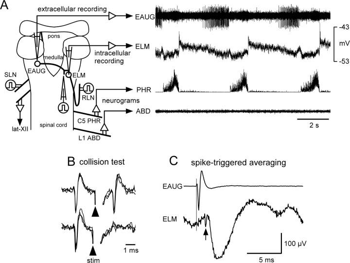
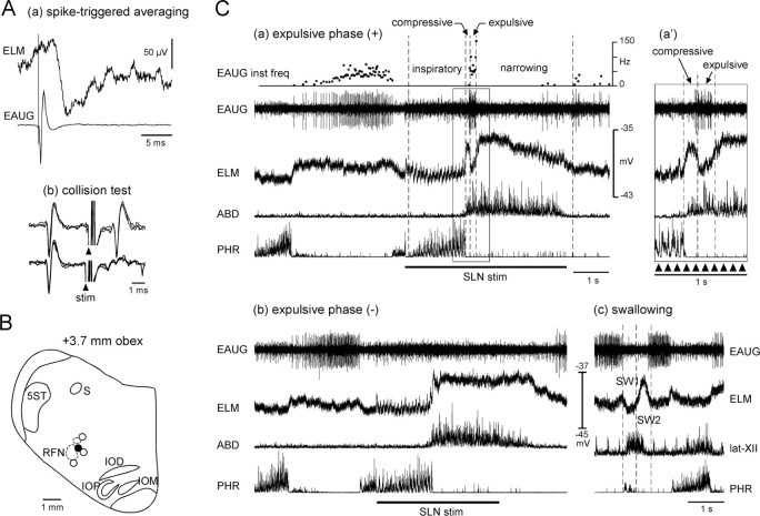
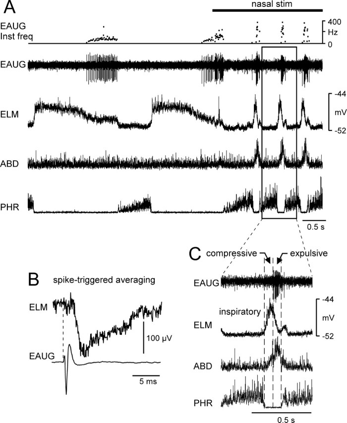
To examine whether motor commands of two or more distinct laryngeal motor patterns converge onto a common premotor network, we conducted dual recordings from the laryngeal adductor motoneuron and its premotor neuron within the brainstem respiratory circuitry during fictive breathing, coughing, sneezing, and swallowing in decerebrate paralyzed cats. Expiratory neurons with an augmenting firing pattern (EAUG), whose action potentials evoked monosynaptic IPSPs in the adductor motoneurons, sharply fired during the expulsive phases of fictive coughing and sneezing, during which the adductor motoneurons transiently repolarized. In contrast, these premotor neurons were silent during the swallow-related hyperpolarization in adductor motoneurons. These results show that one class of medullary respiratory neuron, EAUG, is multifunctional and shared among the central pattern generators (CPGs) for breathing, coughing, and sneezing. In addition, although the CPGs underlying these three behaviors and the swallowing CPG do overlap, EAUG neurons are not part of the swallowing CPG and, in contrast to the other three behaviors, are not a source of inhibitory input to adductor motoneurons during swallowing.


Figures

Figure 1.
Figure 1.
A, Scheme of the dorsal view of the brainstem and spinal cord with the arrangement of the stimulating and recording electrodes used in this experiment. The right panel shows the extracellular spikes of an EAUG neuron in the region spanning the Bötzinger and pre-Bötzinger complexes, the intracellular potential of an ELM, and phrenic (PHR) and abdominal nerve (ABD) activity during normal breathing. lat-XII, Lateral branch of the hypoglossal nerve; RLN, recurrent laryngeal nerve. B, This EAUG neuron was antidromically activated by stimulation in the vicinity of the ELM recording site. Stimulation applied after the spontaneous spikes at less than the critical delay (1.8 ms) resulted in collision of the antidromic spike (3 superimposed sweeps each). C, Monosynaptic IPSP revealed by the spike-triggered averaging method. Top trace, Averaged triggering spike of the EAUG neuron; bottom trace, averaged ELM membrane potential exhibiting hyperpolarizing potential locked to the trigger (470 sweeps). Note a notch indicative of a presynaptic spike in the ELM trace (arrow).
Figure 2.
Figure 2.
Activities of a laryngeal premotor neuron during fictive coughing and swallowing. A, An EAUG neuron-induced monosynaptic IPSP in an ELM. a, Averaged membrane potential of the ELM and averaged triggering spike of the EAUG neuron (824 sweeps). b, Axonal projection of the EAUG neuron to the region of the recorded ELM is examined by the collision test (3 superimposed sweeps each). Arrowheads indicate stimuli applied to the recording site of the ELM. B, Locations of this EAUG neuron (●) and three other EAUG premotor neurons (○). Neurons are projected on a transverse plane at the caudal level of the retrofacial nucleus (RFN) corresponding to the Bötzinger complex. Neurons located at the level of the rostralmost part of the nucleus ambiguus, which corresponds to the pre-Bötzinger complex, are also projected on this plane. The location of the rostralmost nucleus ambiguus is also projected on this plane as a small dotted circle above the retrofacial nucleus. IOD, Dorsal nucleus of the inferior olive; IOM, medial nucleus of the inferior olive; IOP, principal nucleus of the inferior olive; S, solitary tract; 5ST, spinal trigeminal tract. C, Changes in extracellular potential of the EAUG premotor neuron, membrane potential of the ELM, and phrenic (PHR) and abdominal (ABD) nerve activity during fictive coughing and swallowing evoked by SLN stimulation. In this recording, SLN stimulation induced two types of coughing motor patterns: one generated the fictive expulsive phase (a) and the other virtually lacked this phase (b). The period indicated by the rectangular box (a) is expanded on the time axis and shown in the top right corner (a'). When the coughing motor pattern was the former, the ELM hyperpolarized during the fictive inspiratory phase of coughing (labeled as “inspiratory”), depolarized briefly after the termination of the inspiratory phase (the fictive compressive phase labeled as “compressive”), repolarized transiently (the fictive expulsive phase labeled as “expulsive”), and then depolarized again (the fictive narrowing phase labeled as “narrowing”). Periods of SLN stimulation (duration, 0.2 ms; frequency, 10 Hz; intensity, 100 μA) are indicated by horizontal thick lines below PHR recordings or by small arrowheads. Fictive swallowing identified by the hypoglossal burst was also evoked by SLN stimulation (c). In this figure, the neurogram of the lateral branch of the hypoglossal nerve (lat-XII) is shown instead of that of the abdominal nerve. The membrane potential briefly hyperpolarized before the swallow-related hypoglossal burst (SW1) and then abruptly depolarized for the remainder of the hypoglossal burst (SW2). This fictive swallowing was caused during the expiratory phase after cessation of SLN stimulation.
Figure 3.
Figure 3.
A, Intracellular and extracellular recordings from an ELM and its premotor neuron (EAUG) during fictive sneezing. Fictive sneezing is identified by the abdominal burst after phrenic activation during nasal stimulation. The period of stimulation of the nasal mucosa is indicated by a thick horizontal line at the top. B, Spikes of the EAUG neuron-evoked monosynaptic IPSP in the ELM (413 sweeps). C, The period indicated by the rectangular box (A) is expanded on the time axis. This motoneuron hyperpolarized during the inspiratory phase of sneezing and then depolarized abruptly after the termination of the inspiratory phase (the compressive phase). Subsequently, the neuron repolarized during the latter part of the abdominal burst (the expulsive phase). Inst freq, Instantaneous frequency; stim, stimulation; PHR, Phrenic nerve; ABD, abdominal nerve.

Similar articles

Cited by

References

    1. Baekey DM, Morris KF, Gestreau C, Li Z, Lindsey BG, Shannon R. Medullary respiratory neurones and control of laryngeal motoneurones during fictive eupnoea and cough in the cat. J Physiol (Lond) 2001;534:565–581. - PMC - PubMed
    1. Barillot JC, Grélot L, Reddad S, Bianchi AL. Discharge patterns of laryngeal motoneurones in the cat: an intracellular study. Brain Res. 1990;509:99–106. - PubMed
    1. Bartlett DJ. Upper airway motor systems. In: Cherniack NS, Widdicombe JG, editors. Handbook of physiology, Sec 3, The respiratory system. Vol 2. Bethesda, MD: The American Physiological Society; 1986. pp. 223–245.
    1. Batsel HL, Lines AJ. Neural mechanisms of sneeze. Am J Physiol. 1975;229:770–776. - PubMed
    1. Batsel HL, Lines AJ. Discharge of respiratory neurons in sneezes resulting from ethmoidal nerve stimulation. Exp Neurol. 1978;58:410–424. - PubMed

Publication types

MeSH terms

LinkOut - more resources