

Changes in human muscle spindle sensitivity during a proprioceptive attention task
- PMID: 17494703
- PMCID: PMC6672388
- DOI: 10.1523/JNEUROSCI.0572-07.2007
Changes in human muscle spindle sensitivity during a proprioceptive attention task
Abstract
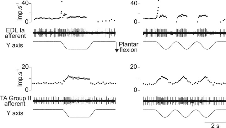
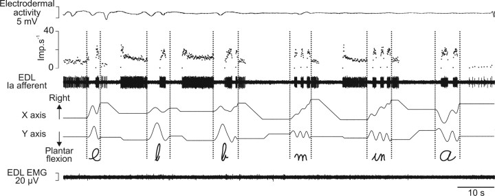
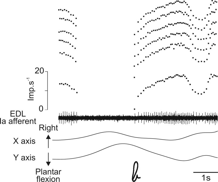
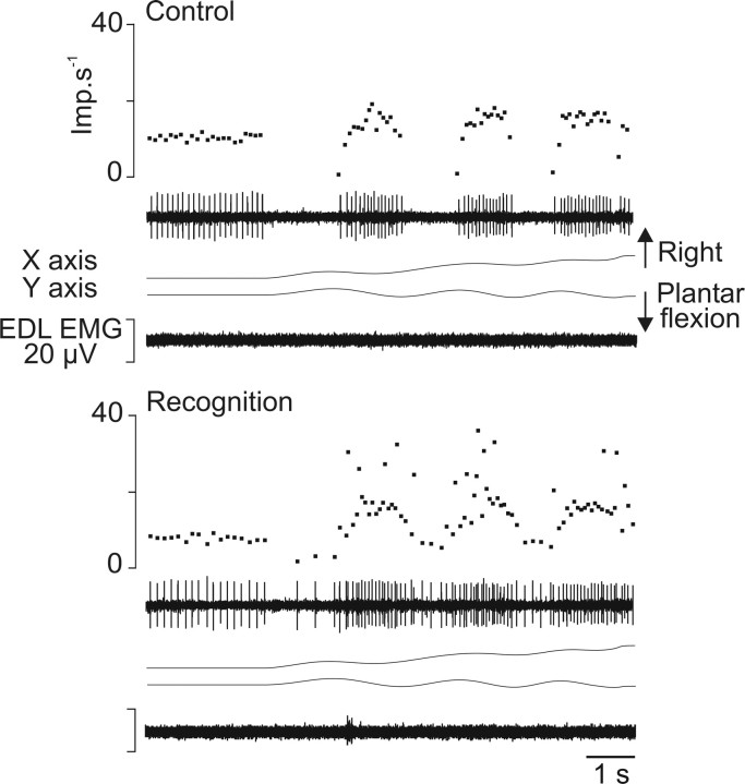
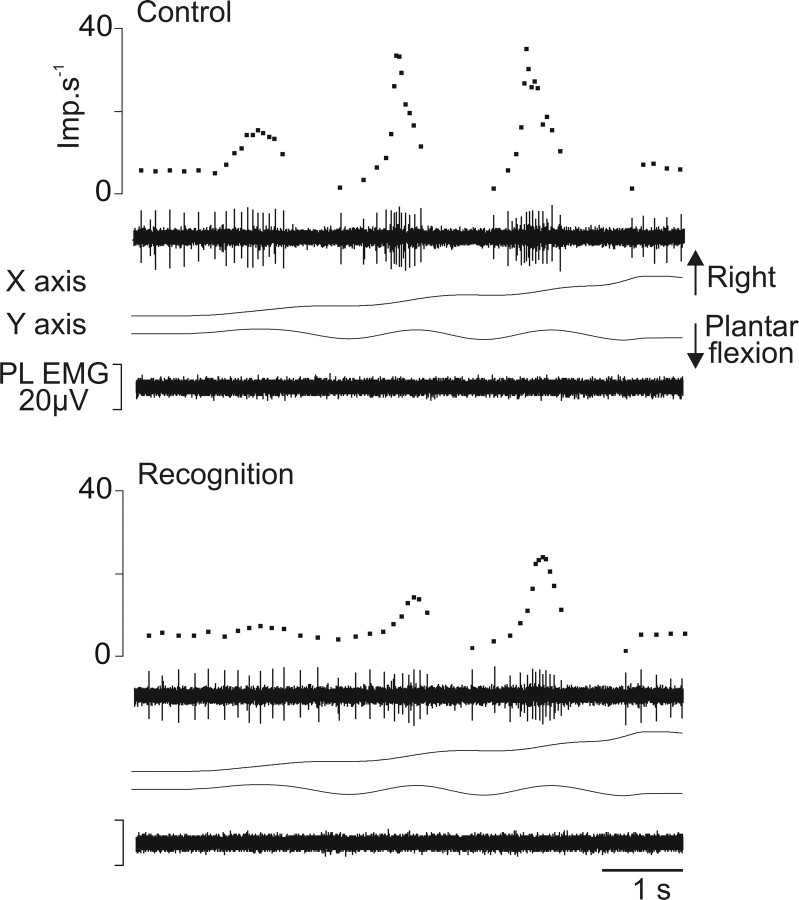
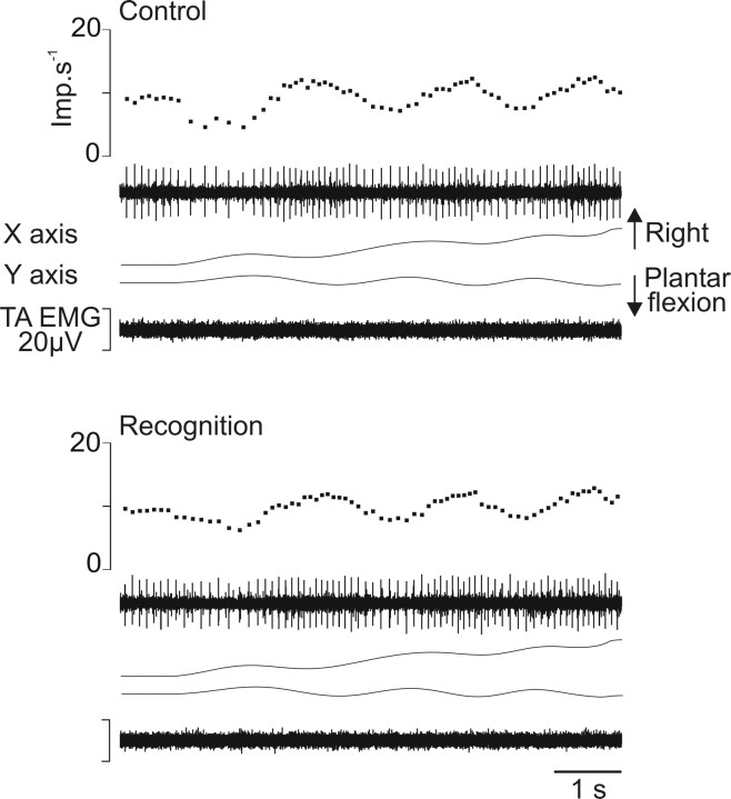
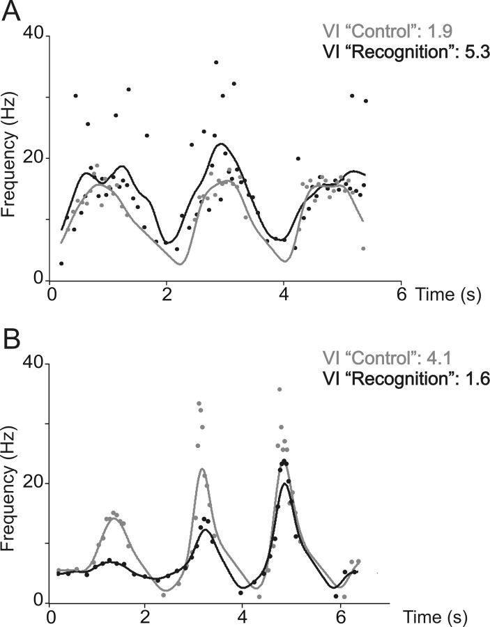
The aim of the present study was to test whether fusimotor control of human muscle spindle sensitivity changed when attention was selectively directed to the recognition of an imposed two-dimensional movement in the form of a written symbol. The unitary activities of 32 muscle spindle afferents (26 Ia, 6 II) were recorded by microneurography at the level of the common peroneal nerve. The patterns of firing rate in response to passive movements of the ankle, forming different letters or numbers, were compared in two conditions: control and recognition. No visual cues were given in either condition, but subjects had to recognize and name the character in one condition compared with not paying attention in the control condition. The results showed that 58% of the tested Ia afferents presented modified responses to movements when these had to be recognized. Changes in Ia afferent responses included decreased depth of modulation, increased variability of discharge, and changes in spontaneous activity. Not all changes were evident in the same afferent. Furthermore, the percentage of correctly recognized movements amounted to 63% when changes were observed, but it was only 48% when the primary ending sensitivity was unaltered. The responses of group II afferents were only weakly changed or unchanged. It is suggested that the altered muscle spindle sensitivity is because of selective changes in fusimotor control, the consequence of which might be to feed the brain movement trajectory information that is more accurate.
Figures
Similar articles
-
"Proprioceptive signature" of cursive writing in humans: a multi-population coding.Exp Brain Res. 2004 Aug;157(3):359-68. doi: 10.1007/s00221-004-1853-x. Epub 2004 Mar 9. Exp Brain Res. 2004. PMID: 15007582
-
Fusimotor drive may adjust muscle spindle feedback to task requirements in humans.J Neurophysiol. 2009 Feb;101(2):633-40. doi: 10.1152/jn.91041.2008. Epub 2008 Nov 26. J Neurophysiol. 2009. PMID: 19036863
-
Proprioceptive feedback in humans expresses motor invariants during writing.Exp Brain Res. 2005 Jul;164(2):242-9. doi: 10.1007/s00221-005-2246-5. Epub 2005 Apr 27. Exp Brain Res. 2005. PMID: 15856208
-
Fusimotor control of proprioceptive feedback during locomotion and balancing: can simple lessons be learned for artificial control of gait?Prog Brain Res. 1993;97:173-80. doi: 10.1016/s0079-6123(08)62275-x. Prog Brain Res. 1993. PMID: 8234743 Review.
-
What is the role of muscle receptors in proprioception?Muscle Nerve. 2005 Jun;31(6):780-7. doi: 10.1002/mus.20330. Muscle Nerve. 2005. PMID: 15818635 Review.
Cited by
-
Noise-enhanced kinaesthesia: a psychophysical and microneurographic study.Exp Brain Res. 2013 Aug;228(4):503-11. doi: 10.1007/s00221-013-3581-6. Epub 2013 May 28. Exp Brain Res. 2013. PMID: 23712687 Clinical Trial.
-
Vibrotactile cuing revisited to reveal a possible challenge to sensorimotor adaptation.Exp Brain Res. 2016 Dec;234(12):3523-3530. doi: 10.1007/s00221-016-4750-1. Epub 2016 Aug 8. Exp Brain Res. 2016. PMID: 27501732
-
Emotions alter muscle proprioceptive coding of movements in humans.Sci Rep. 2017 Aug 16;7(1):8465. doi: 10.1038/s41598-017-08721-4. Sci Rep. 2017. PMID: 28814736 Free PMC article.
-
Can proprioceptive training improve motor learning?J Neurophysiol. 2012 Dec;108(12):3313-21. doi: 10.1152/jn.00122.2012. Epub 2012 Sep 12. J Neurophysiol. 2012. PMID: 22972960 Free PMC article. Clinical Trial.
-
The Dynamics of Voluntary Force Production in Afferented Muscle Influence Involuntary Tremor.Front Comput Neurosci. 2016 Aug 19;10:86. doi: 10.3389/fncom.2016.00086. eCollection 2016. Front Comput Neurosci. 2016. PMID: 27594832 Free PMC article.
References
-
- Albert F, Ribot-Ciscar E, Fiocchi M, Bergenheim M, Roll JP. Proprioceptive feedback in humans expresses motor invariants during writing. Exp Brain Res. 2005;164:242–249. - PubMed
-
- Bergenheim M, Johansson H, Pedersen J. The role of the gamma-system for improving information transmission in populations of Ia afferents. Neurosci Res. 1995;23:207–215. - PubMed
-
- Bergenheim M, Roll JP, Ribot-Ciscar E. Microneurography in humans. In: Windhorst U, Johansson H, editors. Modern techniques in neuroscience research. New York: Springer; 1999. pp. 801–819.
-
- Bergenheim M, Ribot-Ciscar E, Roll JP. Proprioceptive population coding of two-dimensional limb movements in humans: I. Muscle spindle feedback during spatially oriented movements. Exp Brain Res. 2000;134:301–310. - PubMed
-
- Celichowski J, Emonet-Denand F, Laporte Y, Petit J. Distribution of static gamma axons in cat peroneus tertius spindles determined by exclusively physiological criteria. J Neurophysiol. 1994;71:722–732. - PubMed
Publication types
MeSH terms
LinkOut - more resources
Full Text Sources
Medical