. 2007 May 9;27(19):5179-89.
doi: 10.1523/JNEUROSCI.5499-06.2007.

TRPC3 channels are necessary for brain-derived neurotrophic factor to activate a nonselective cationic current and to induce dendritic spine formation

Affiliations

TRPC3 channels are necessary for brain-derived neurotrophic factor to activate a nonselective cationic current and to induce dendritic spine formation

Michelle D Amaral et al. J Neurosci. .

Abstract

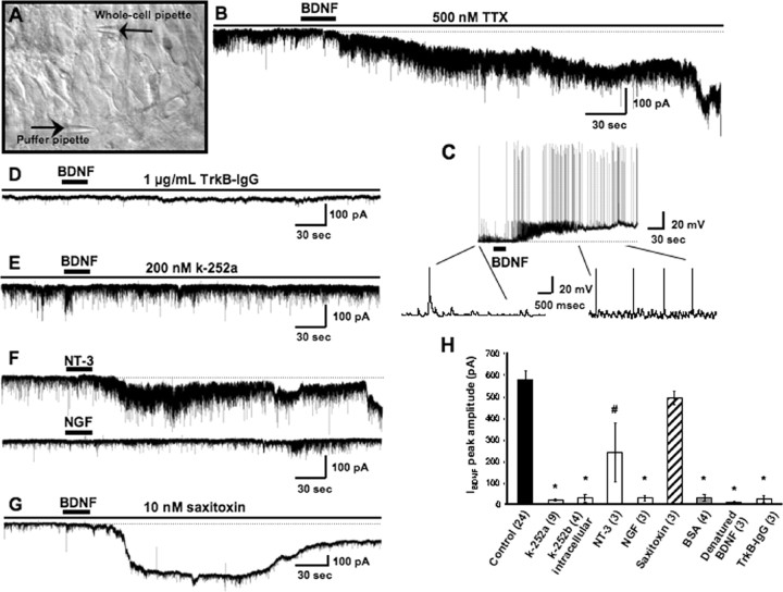
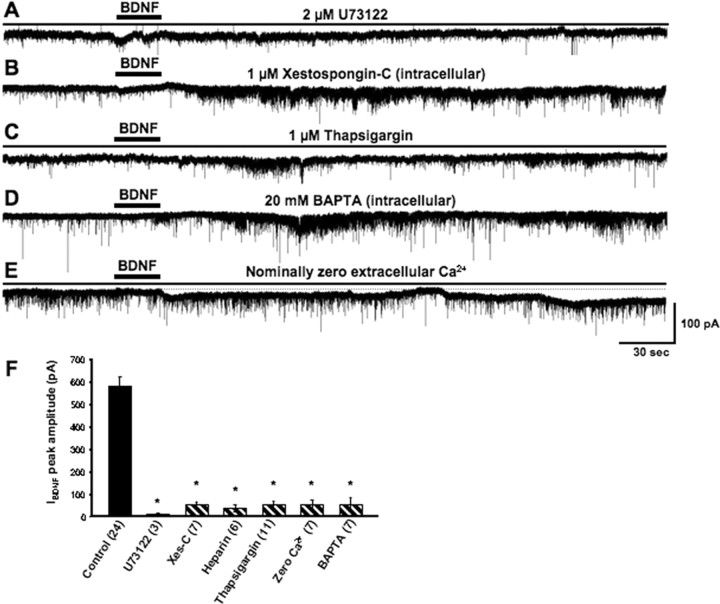
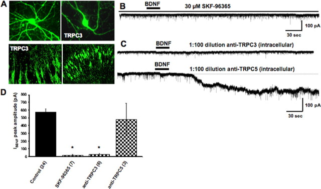
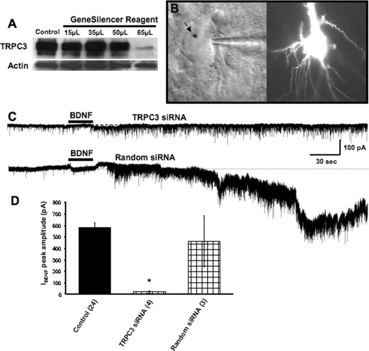
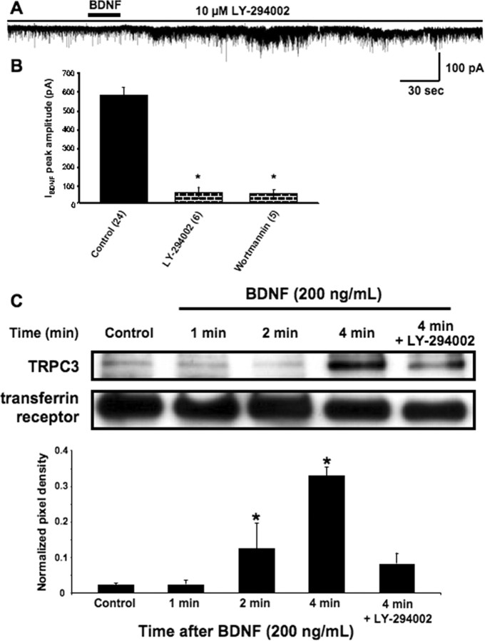
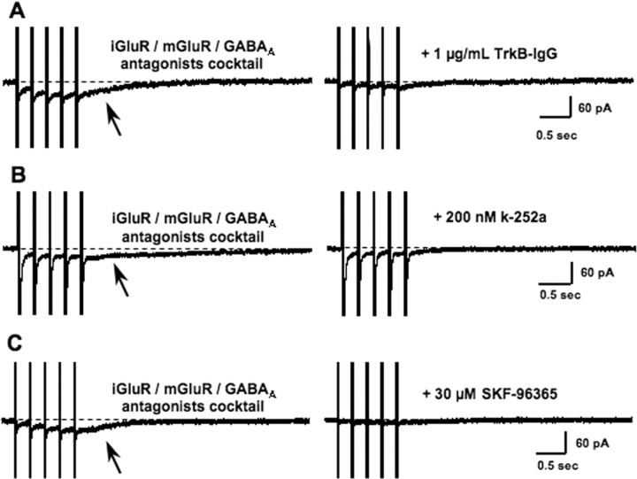
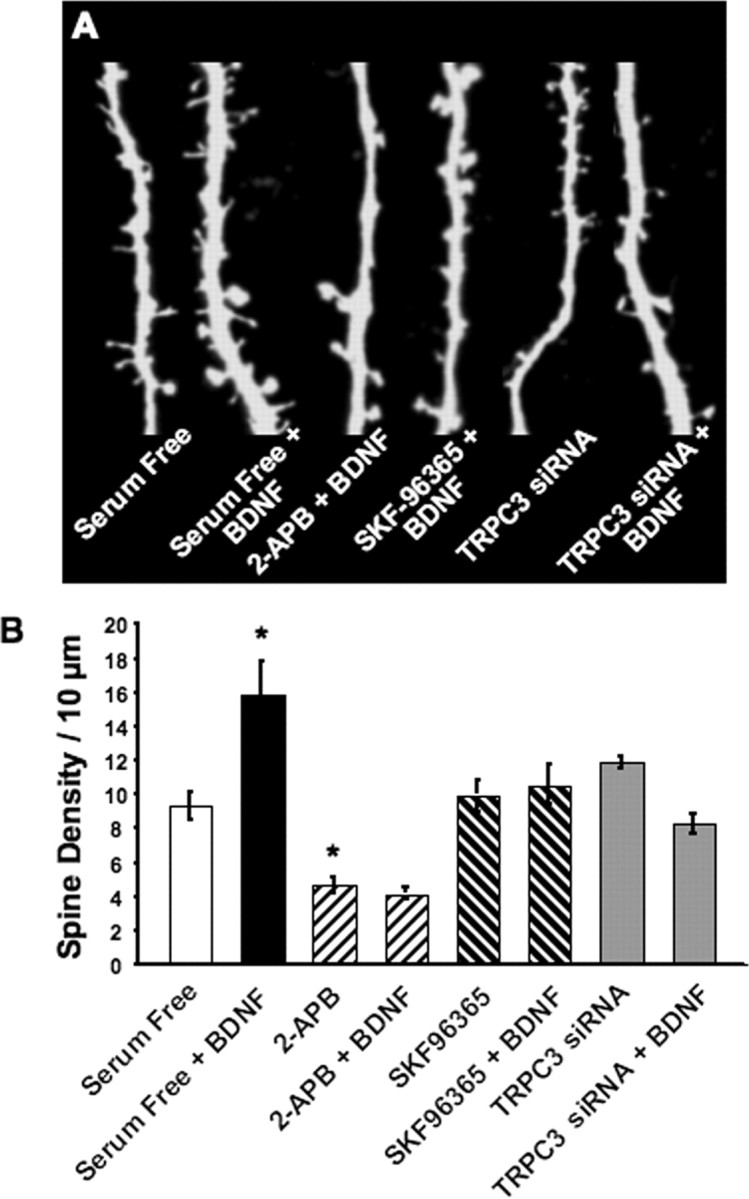
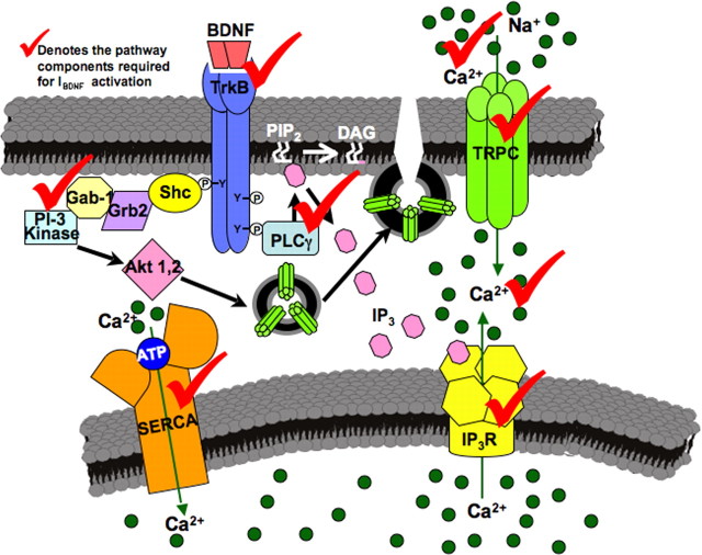
Brain-derived neurotrophic factor (BDNF) exerts prominent effects on hippocampal neurons, but the mechanisms that initiate its actions are poorly understood. We report here that BDNF evokes a slowly developing and sustained nonselective cationic current (I(BDNF)) in CA1 pyramidal neurons. These responses require phospholipase C, IP3 receptors, Ca2+ stores, and Ca2+ influx, suggesting the involvement of transient receptor potential canonical subfamily (TRPC) channels. Indeed, I(BDNF) is absent after small interfering RNA-mediated TRPC3 knockdown. The sustained kinetics of I(BDNF) appears to depend on phosphatidylinositol 3-kinase-mediated TRPC3 membrane insertion, as shown by surface biotinylation assays. Slowly emerging membrane currents after theta burst stimulation are sensitive to the scavenger TrkB-IgG and TRPC inhibitors, suggesting I(BDNF) activation by evoked released of endogenous, native BDNF. Last, TRPC3 channels are necessary for BDNF to increase dendritic spine density. Thus, TRPC channels emerge as novel mediators of BDNF-mediated dendritic remodeling through the activation of a slowly developing and sustained membrane depolarization.


Figures

Figure 1.
Figure 1.
BDNF activation of TrkB receptors induces a slowly developing and sustained depolarizing membrane response that is not mediated by STX-sensitive Na+ channels. A, Representative hippocampal slice culture (from a postnatal day rat, 10 div) imaged by IR-DIC showing a whole-cell electrode patched on a CA1 neuron and the position of the BDNF application pipette. For clarity, the BDNF pipette has been lowered to the slice surface to show its position relative to the whole-cell electrode. B, Representative example of IBDNF, the membrane current evoked by BDNF (100 μg/ml in the puffer pipette) in a CA1 pyramidal neuron voltage clamped near its resting membrane potential (Vh of − 65 mV; Cs-gluconate electrode). C, Under current clamp, BDNF induces a membrane depolarization with similar kinetics to that of IBDNF and increases spontaneous synaptic activity and spike frequency (K-gluconate electrode, no TTX). D, The selective BDNF scavenger TrkB–IgG (1 μg/ml) prevented the responses to the first two to three exogenous BDNF applications, suggesting that the effective BDNF concentration necessary to activate IBDNF is between 33 and 50 ng/ml, equivalent to 1.2–1.8 nm of the BDNF dimer, well within the TrkB receptor affinity. E, The tyrosine kinase inhibitor k-252a (200 nm) prevented the activation of IBDNF. F, NT-3 induced a membrane current of similar kinetics but smaller amplitude than IBDNF (top), whereas NGF did not affect membrane conductance (bottom; both neurotrophins at 100 μg/ml in the application pipette). G, Contrary to the fast Na+ current mediated by Nav1.9 channels, IBDNF is insensitive to STX (10 nm). H, Average amplitude of IBDNF in the above experimental conditions (*p < 0.0001, #p < 0.005 vs BDNF application in TTX).
Figure 2.
Figure 2.
IBDNF activation requires PLC activity, functional IP3 receptors, full intracellular stores, Ca2+ influx, and intracellular Ca2+ elevations. A, Bath application of the PLC inhibitor U73122 completely blocked IBDNF. B, Intracellular application of xestospongin-C (Xes-C; 1 μm), a specific inhibitor of IP3Rs, prevents IBDNF. C, Depletion of intracellular stores with the SERCA pump inhibitor thapsigargin (1 μm) also abolishes IBDNF. D, Consistent with a requirement of Ca2+ signaling, IBDNF was also inhibited by loading neurons with the fast Ca2+ chelator BAPTA (20 mm). E, Activation of IBDNF also requires extracellular Ca2+. F, Average amplitude of IBDNF in the above experimental conditions (*p < 0.0001 vs BDNF application in TTX).
Figure 3.
Figure 3.
IBDNF is sensitive to inhibitors of TRPC currents, which are expressed in hippocampal neurons. A, TRPC3 localized in the cell body and dendritic processes of pyramidal-like hippocampal neurons in primary culture (top). TRPC3 was also detected in neuronal cell bodies and dendritic processes of the CA1 region of cultured slices (bottom). B, The TRPC/SOC inhibitor SKF-96365 (30 μm) prevents IBDNF. C, Antibodies against intracellular domains of TRPC3 included in the whole-cell recording solution completely abolish IBDNF (top), whereas anti-TRPC5 had no effect (bottom). D, Average amplitude of IBDNF in the above experimental conditions (*p < 0.0001 vs BDNF application in TTX).
Figure 4.
Figure 4.
TRPC3 subunits are necessary for the activation of IBDNF. A, siRNA designed to knockdown TRPC3 expression reduced TRPC3 protein levels in primary cultured neurons. B, IR-DIC image (left) and eYFP fluorescence (488 nm excitation) of a CA1 pyramidal neuron biolistically transfected with siRNA oligos and eYFP cDNA. Note the gold particle (arrow) on the nucleus of the neuron and the whole-cell patch pipette toward its right. C, BDNF failed to induce a membrane current in CA1 pyramidal neurons transfected with TRPC3 siRNA (top), whereas biolistic transfection with random siRNA oligos and eYFP cDNA does not affect IBDNF amplitude or activation kinetics. D, Average amplitude of IBDNF in the above experimental conditions (*p < 0.0001 vs BDNF application in TTX).
Figure 5.
Figure 5.
PI3 kinase signaling is required for IBDNF and the rapid increase in TRPC3 surface content. A, IBDNF was reduced by the PI3 kinase inhibitor LY-294002 (10 μm). B, Average amplitude of IBDNF in the above experimental conditions (*p < 0.0001 vs BDNF application in TTX). C, Surface biotinylation reactions on cultured hippocampal neurons revealed that levels of surface accessible TRPC3 progressively increased after BDNF application with a time course that parallels the time course of IBDNF activation (1, 2, and 4 min). In addition, the same PI3 kinase inhibitor that reduced IBDNF also prevented the surface translocation of TRPC3. Bar graphs shows the quantitative analysis of surface biotinylation reactions, using the transferrin receptor for normalization of pixel intensity.
Figure 6.
Figure 6.
A, Representative examples of membrane currents evoked by TBS in the presence of ionotropic and metabotropic glutamate and GABAA receptors outlasted the afferent train (arrows). These slow currents were completely blocked by TrkB–IgG (1 μg/ml). B, Slow currents evoked by TBS were completely blocked by k-252a (200 nm). C, TBS-evoked slow currents were completely blocked by SKF-96365 (30 μm).
Figure 7.
Figure 7.
Functional TRPC3 channels are required for BDNF to increase dendritic spine density in CA1 pyramidal neurons. Slice cultures were transfected with either eYFP cDNA alone or in combination with TRPC3 siRNA oligonucleotides. Slices were exposed to BDNF (200 ng/ml in serum-free media) for 24 h, and spine density was measured by confocal microscopy 24 h after transfection. A, Representative examples of segments of apical secondary and tertiary dendritic branches of eYFP-transfected CA1 pyramidal neurons. The TRPC inhibitors SKF-96365 and 2-APB prevented the increase in spine density by BDNF. Likewise, siRNA-mediated knockdown of TRPC3 abolished the effect of BDNF on spine formation, without effects of its own. B, Average spine density in the above mentioned experimental groups. *p < 0.05 compared with serum-free controls, as per ANOVA followed by Newman–Keuls multiple comparison post hoc test.
Figure 8.
Figure 8.
Schematic representation of the proposed model for intracellular signaling cascades initiated by BDNF stimulation of TrkB receptors leading to the activation of membrane currents and Ca2+ signals mediated by TRPC3-containing channels. Gab-1, Growth-associated binder 1; Grb2, growth factor receptor-bound protein 2; Shc, Src homology 2 domain-containing transforming protein C1; P, phosphate groups; Y, tyrosine residues.

Similar articles

Cited by

References

    1. Agell N, Bachs O, Rocamora N, Villalonga P. Modulation of the Ras/Raf/MEK/ERK pathway by Ca2+, and calmodulin. Cell Signal. 2002;14:649–654. - PubMed
    1. Alonso M, Medina JH, Pozzo-Miller L. ERK1/2 activation is necessary for BDNF to increase dendritic spine density in hippocampal CA1 pyramidal neurons. Learn Mem. 2004;11:172–178. - PMC - PubMed
    1. Alzheimer C. A novel voltage-dependent cation current in rat neocortical neurones. J Physiol (Lond) 1994;479:199–205. - PMC - PubMed
    1. Amaral MD, Chapleau CA, Pozzo-Miller L. Transient receptor potential channels as novel effectors of brain-derived neurotrophic factor signaling: potential implications for Rett syndrome. Pharmacol Ther. 2007;113:394–409. - PMC - PubMed
    1. Balkowiec A, Katz DM. Cellular mechanisms regulating activity-dependent release of native brain-derived neurotrophic factor from hippocampal neurons. J Neurosci. 2002;22:10399–10407. - PMC - PubMed

Publication types

MeSH terms

LinkOut - more resources