. 2007 May 15;104(20):8449-54.
doi: 10.1073/pnas.0703039104. Epub 2007 May 9.

Hepatitis C virus epitope-specific neutralizing antibodies in Igs prepared from human plasma

Affiliations

Hepatitis C virus epitope-specific neutralizing antibodies in Igs prepared from human plasma

Pei Zhang et al. Proc Natl Acad Sci U S A. .

Abstract

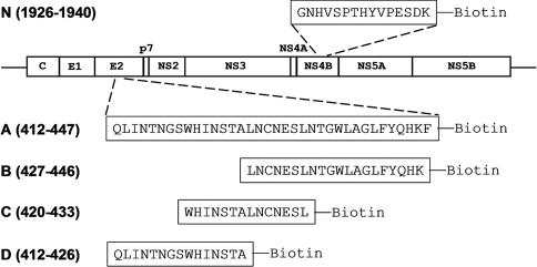
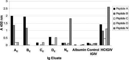
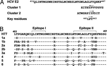
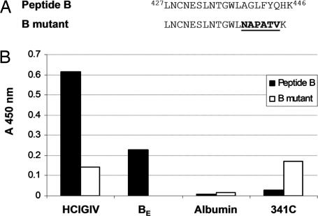
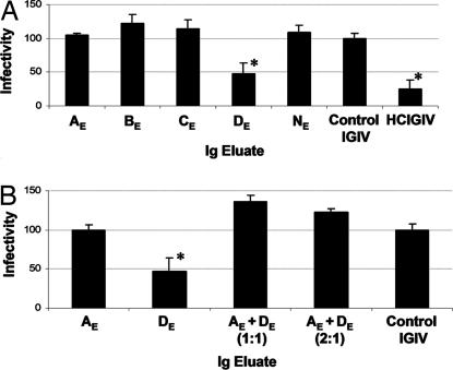
Neutralizing antibodies directed against hepatitis C virus (HCV) are present in Igs made from anti-HCV-positive plasma. However, these HCV-specific Igs are largely ineffective in vivo. The mechanism for the poor effectiveness is currently unknown. We hypothesize that the presence of nonneutralizing antibodies in HCV-specific Igs interferes with the function of neutralizing antibodies, resulting in the reduction or blockage of their effect. In the present study, we identified at least two epitopes at amino acid residues 412-419 (epitope I) and 434-446 (epitope II), located downstream of the hypervariable region I within the HCV E2 protein. We demonstrated that epitope I, but not epitope II, was implicated in HCV neutralization and that binding of a nonneutralizing antibody to epitope II completely disrupted virus neutralization mediated by antibody binding at epitope I. The dynamic interaction between nonneutralizing and neutralizing antibodies may thus play a key role in determining the outcomes of HCV infection. Further exploration of this interplay should lead to a better understanding of the mechanisms of neutralization and immune escape and may indicate pathways for the manufacture of an effective HCV-specific Ig product for immune prophylaxis of HCV infection.


Conflict of interest statement

The authors declare no conflict of interest.

Figures

Fig. 1.
Fig. 1.
Schematic representation of HCV polyprotein and biotin-linked peptides (A, B, C, D, and N) used in this study. All peptides were synthesized based on the sequence of strain H77. The numbers indicate the position of these peptides in HCV polyprotein, and single letters are used to represent amino acids.
Fig. 2.
Fig. 2.
Presence of HCV E2 peptide-specific antibodies in HCIGIV. (A) Determination of HCIGIV antibody titer to peptide A. The x axis indicates the dilution of HCIGIV, and the y axis indicates absorbance at 450 nm in ELISA. Albumin (5%) and a control IGIV (5%) at 1:400 dilution in PBS were used as controls. (B) Spiking of HCIGIV into the control IGIV. The x axis indicates dilution of HCIGIV in the control IGIV. HCIGIV at 1:400 dilution alone was used as the positive control. Albumin (5%) and the control IGIV (5%) at 1:400 dilution were used as negative controls. The y axis indicates absorbance at 450 nm in ELISA.
Fig. 3.
Fig. 3.
Determination of HCV epitope-specific antibodies in HCIGIV. The x axis indicates Ig eluates (AE, BE, CE, DE, or NE) collected after affinity binding and elution of HCIGIV by using a given peptide (peptide A, B, C, D, or N). HCIGIV at 1:400 dilution alone was used as the positive control. Albumin (5%) and the control IGIV (5%) at 1:400 dilution were used as negative controls. The y axis indicates absorbance at 450 nm in ELISA, representing specific binding of a given Ig eluate to each individual peptide.
Fig. 4.
Fig. 4.
Summary of antibody binding and location of epitopes. (A) Antibody-binding activity in Ig eluates for individual peptides. The data from Fig. 3 are summarized. >, stronger than; =, equal to; −, no detectable peptide binding. (B) Identification of epitopes within HCV E2 protein. The sequences of the identified epitopes are underlined.
Fig. 5.
Fig. 5.
Epitope mapping. (A) HCV epitope mapping by screening a random peptide phage-display library. Amino acid sequences of two phage clusters identified by screening a phage-display library (PhD-12) with AE as a source of antibodies are indicated. The direction of the arrow indicates the arrangement of the residues in the peptide critical for antibody binding. The key residues for the epitope are numbered based on the amino acid sequence of strain H77. (B) Alignment of amino acid sequences of the E2 regions among various HCV genotypes (10, 22). Epitopes I and II identified in this study are shown. A hyphen indicates an amino acid residue identical to that of the H77 sequence.
Fig. 6.
Fig. 6.
Identification of HCV epitope by mutation analysis. (A) Mutation of epitope II. Amino acid sequences for peptide B and its mutation (B mutant) are presented. The mutation site is underlined. (B) Detection of antibody binding by ELISA. A total of 100 ng of biotin-conjugated peptide B and its mutant (B mutant) were added to streptavidin-coated 96-well plates in an ELISA. The x axis indicates antibodies that were used in this assay. HCIGIV at 1:800 dilution was used as the positive control, and albumin (5%) at 1:800 dilution was used as the negative control. 341C, a monoclonal antibody that recognizes the sequence NAPATV, was used at 1:200 dilution. The y axis indicates absorbance at 450 nm, representing specific binding of a given antibody to each individual peptide.
Fig. 7.
Fig. 7.
HCV neutralization in cell culture. (A) HCV neutralization by Ig eluates. The x axis indicates Ig eluates that were used in this assay at 1:40 dilution. HCIGIV at 1:100 dilution was used as the positive control, and an IGIV (5%) at 1:100 dilution was used as the negative control. The y axis indicates infectivity (percentage of negative control). The asterisk indicates statistical significance (P < 0.05). (B) Blocking neutralizing activity of DE by nonneutralizing Ig in AE. The x axis indicates Ig AE or DE alone at 1:40 dilution or a mixture of AE and DE (AE + DE) at 1:1 or 2:1 ratio. An IGIV (5%) at 1:100 dilution was used as the negative control. The y axis indicates infectivity (percentage of negative control). The asterisk indicates statistical significance (P < 0.05).

References

    1. Alter HJ, Seeff LB. Semin Liver Dis. 2000;20:17–35. - PubMed
    1. Bartosch B, Dubuisson J, Cosset FL. J Exp Med. 2003;197:633–642. - PMC - PubMed
    1. Thimme R, Bukh J, Spangenberg HC, Wieland S, Pemberton J, Steiger C, Govindarajan S, Purcell RH, Chisari FV. Hepatology. 2003;37:1472–1474. - PubMed
    1. Bartosch B, Bukh J, Meunier JC, Granier C, Engle RE, Blackwelder WC, Emerson SU, Cosset FL, Purcell RH. Proc Natl Acad Sci USA. 2003;100:14199–14204. - PMC - PubMed
    1. Meunier JC, Engle RE, Faulk K, Zhao M, Bartosch B, Alter H, Emerson SU, Cosset FL, Purcell RH, Bukh J. Proc Natl Acad Sci USA. 2005;102:4560–4565. - PMC - PubMed

Publication types

LinkOut - more resources