. 2007 May 15;104(20):8508-13.
doi: 10.1073/pnas.0611391104. Epub 2007 May 9.

ApoER2/VLDL receptor and Dab1 in the rostral migratory stream function in postnatal neuronal migration independently of Reelin

Affiliations

ApoER2/VLDL receptor and Dab1 in the rostral migratory stream function in postnatal neuronal migration independently of Reelin

Nuno Andrade et al. Proc Natl Acad Sci U S A. .

Abstract

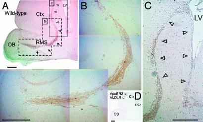
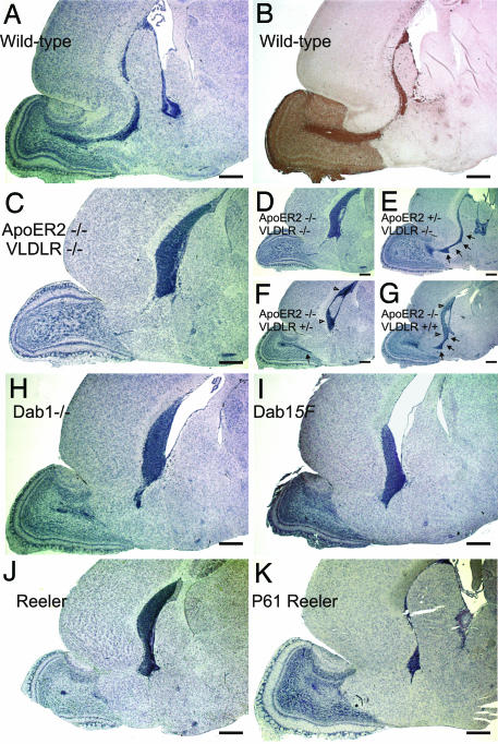
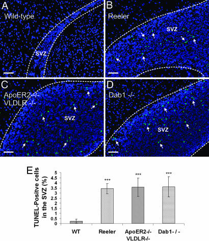
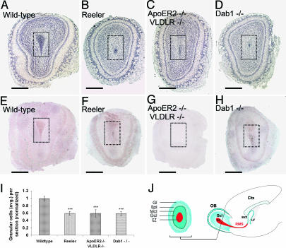
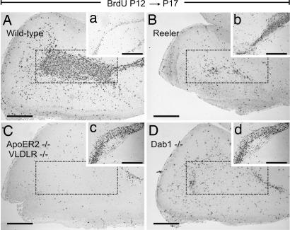
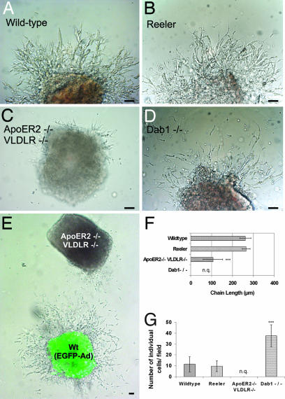
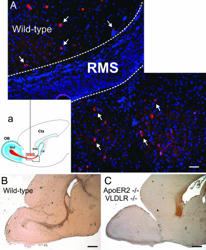
Postnatal migration of interneuron precursors from the subventricular zone to the olfactory bulb occurs in chains that form the substrate for the rostral migratory stream. Reelin is suggested to induce detachment of neuroblasts from the chains when they arrive at the olfactory bulb. Here we show that ApoER2 and possibly very-low-density lipoprotein receptor (VLDLR) and their intracellular adapter protein Dab1 are involved in chain formation most likely independent of Reelin. F-spondin, which is present in the stream, may act as ligand for ApoER2 and VLDLR. In mice lacking either both receptors or Dab1 chain formation is severely compromised, and as a consequence the rostral migratory stream is virtually absent and neuroblasts accumulate in the subventricular zone. The mutant animals exhibit severe neuroanatomical defects in the subventricular zone and in the olfactory bulb. These data demonstrate a cell-autonomous function of ApoER2, and most likely VLDLR and Dab1, in postnatal migration of neuroblasts in the forebrain, which is suggested to depend on ligands other than Reelin.


Conflict of interest statement

The authors declare no conflict of interest.

Figures

Fig. 1.
Fig. 1.
Neuroblasts migrating within the RMS express ApoER2. Sagittal sections (5 μm) of the forebrains from WT mice (A–C) and from ApoER2−/−/VLDLR−/− mice (D) were immunostained with a polyclonal antibody against ApoER2. The RMS is highlighted by arrows, and the SVZ is highlighted by open arrowheads. Ctx, cortex; LV, lateral ventricle; St, striatum. (Scale bars: 500 μm.)
Fig. 2.
Fig. 2.
Ablation of critical components of the Reelin signaling pathway leads to loss of the RMS and accumulation of neuroblasts in the SVZ. Sagittal sections (5 μm) of the forebrains of WT (A and B), ApoER2−/−/VLDLR−/− (C and D), ApoER2+/−/VLDLR−/− (E), ApoER2−/−/VLDLR+/− (F), ApoER2−/−/VLDLR+/+ (G), Dab1−/− (H), Dab1-5F (I), and reeler (J) mice at P17 and reeler mice (K) at P61 were stained with hematoxylin (A and C–K) or with an antibody against doublecortin (B). Sections were taken from three to five independent animals from each of the genetic backgrounds, and representative sections are displayed. (Scale bars: 500 μm.) Arrows are used to highlight the RMS.
Fig. 3.
Fig. 3.
Cell accumulation in the SVZs of reeler, ApoER2−/−/VLDLR−/−, and Dab1−/− mice leads to increased apoptosis. Sagittal sections (5 μm) prepared from the forebrains of WT (A), reeler (B), ApoER2−/−/VLDLR−/− (C), and Dab1−/− (D) mice were analyzed for the presence of apoptotic cells by TUNEL assays. (E) Apoptosis was quantified based on the percentage of TUNEL-positive cells in the SVZ using five to eight matched 5-μm sections from three animals from each genetic background. Plots show average ± SEM. ∗∗∗, P < 0.0005 (Student's t test). (Scale bars: 50 μm.)
Fig. 4.
Fig. 4.
OBs from reeler mice, ApoER2−/−/VLDLR−/− mice, and Dab1−/− mice have a reduced or vestigial EZ. Matched coronal sections (5 μm) derived from the OBs of WT mice (A and E), reeler mice (B and F), ApoER2−/−/VLDLR−/− mice (C and G), and Dab1−/− mice (D and H) were stained with hematoxylin (A–D) or immunostained for ApoER2 (E–H). The EZ (darker area inside the doted squares) is drastically reduced in the mutant OBs because of the disruption of the corresponding RMS (B–D). (I) Quantification of the cell numbers present in the Gcl and EZ (normalized to the cell number in the Gcl of WT mice) displayed a 40% decrease in the mutant OBs. Average cell numbers were calculated from 11 matched sections from two WT mice, 19 matched sections from three reeler mice, 18 matched sections from three ApoER2−/−/VLDLR−/− mice, and 18 matched sections from three Dab1−/− mice. Plots show average ± SEM. ∗∗∗, P < 0.0005 (Student's t test). (J) Schematic illustration of a coronal section from an OB (Left) and a sagittal section of a forebrain (Right). (Scale bars: 500 μm.) Ctx, cortex; LV, lateral ventricle.
Fig. 5.
Fig. 5.
Migration of newly generated neuroblasts is affected in the forebrains of mice carrying mutations in the Reelin signaling pathway. WT (A), reeler (B), ApoER2−/−/VLDLR−/− (C), and Dab1−/− (D) mice received a BrdU pulse at P12, and brains were collected at P17. Matched sagittal sections of the forebrains were stained for BrdU-positive cells. Areas corresponding to the EZs are marked by dotted frames. (a–d) BrdU staining of the corresponding SVZs. (Scale bars: 500 μm.)
Fig. 6.
Fig. 6.
Chain formation from explants of the SVZ is normal in reeler but severely disturbed in ApoER2−/−/VLDLR−/− and Dab1−/− mice. SVZ explants were prepared from P5 WT (A), reeler (B), ApoER2−/−/VLDLR−/− (C), and Dab1−/− (D) mice and analyzed after 50 h in culture by measuring the length of migratory chains (F, distance between the bases to the tips of the chains) and the number of individual cells per field (G). For quantification 19 WT, 16 reeler, 20 ApoER2−/−/VLDLR−/−, and 21 Dab1−/− explants from two mice of each phenotype were used. Plots show average ± SEM. ∗∗∗, P < 0.0001 (Student's t test). (E) WT SVZ explants infected with EGFP adenovirus were cocultured with explants from ApoER2−/−/VLDLR−/− mice. n.q., not quantified. (Scale bar: 50 μm.)
Fig. 7.
Fig. 7.
Expression of Reelin and F-spondin in the RMS. (A) Reelin-positive cells accompany the RMS. Sagittal sections (5 μm) prepared from the forebrains of WT mice were immunostained with an antibody against Reelin. The RMS is highlighted by dotted lines. Reelin-positive cells are indicated by arrows. (a) Schematic representation of the area shown in A. (Scale bar: 50 μm.) (B and C) F-spondin is present in the RMS and accumulates in the SVZ of ApoER2−/−/VLDLR−/− mice. Sagittal sections (5 μm) prepared from the forebrains of WT (B) and ApoER2−/−/VLDLR−/− (C) mice were immunostained with an antibody against F-spondin. (Scale bar: 500 μm.)

Similar articles

Cited by

References

    1. Luskin MB. Neuron. 1993;11:173–189. - PubMed
    1. Lois C, Alvarez-Buylla A. Science. 1994;264:1145–1148. - PubMed
    1. Lois C, Garcia-Verdugo JM, Alvarez-Buylla A. Science. 1996;271:978–981. - PubMed
    1. Wichterle H, Garcia-Verdugo JM, Alvarez-Buylla A. Neuron. 1997;18:779–791. - PubMed
    1. Alberti S, Krause SM, Kretz O, Philippar U, Lemberger T, Casanova E, Wiebel FF, Schwarz H, Frotscher M, Schutz G, Nordheim A. Proc Natl Acad Sci USA. 2005;102:6148–6153. - PMC - PubMed

Publication types

MeSH terms