. 2007 Jul;18(7):2646-55.
doi: 10.1091/mbc.e06-10-0897. Epub 2007 May 9.

PI4P promotes the recruitment of the GGA adaptor proteins to the trans-Golgi network and regulates their recognition of the ubiquitin sorting signal

Affiliations

PI4P promotes the recruitment of the GGA adaptor proteins to the trans-Golgi network and regulates their recognition of the ubiquitin sorting signal

Jing Wang et al. Mol Biol Cell. 2007 Jul.

Abstract

Phosphatidylinositol 4 phosphate (PI4P) is highly enriched in the trans-Golgi network (TGN). Here we establish that PI4P is a key regulator of the recruitment of the GGA clathrin adaptor proteins to the TGN and that PI4P has a novel role in promoting their recognition of the ubiquitin (Ub) sorting signal. Knockdown of PI4KIIalpha by RNA interference (RNAi), which depletes the TGN's PI4P, impaired the recruitment of the GGAs to the TGN. GGAs bind PI4P primarily through their GAT domain, in a region called C-GAT, which also binds Ub but not Arf1. We identified two basic residues in the GAT domain that are essential for PI4P binding in vitro and for the recruitment of GGAs to the TGN in vivo. Unlike wild-type GGA, GGA with mutated GATs failed to rescue the abnormal TGN phenotype of the GGA RNAi-depleted cells. These residues partially overlap with those that bind Ub, and PI4P increased the affinity of the GAT domain for Ub. Because the recruitment of clathrin adaptors and their cargoes to the TGN is mediated through a web of low-affinity interactions, our results show that the dual roles of PI4P can promote specific GGA targeting and cargo recognition at the TGN.


Figures
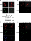
Figure 1.
Figure 1.
Effects of PI4KIIα RNAi on the intracellular distribution of the GGAs and Arf in cells. (A–C) Effects on GGA localization. Endogenous GGA1, 2, and 3 were detected with isoform-specific antibodies. The TGN was stained with anti-TGN46. Western blot in A shows the partitioning of GGA1, PI4KIIα, and βCOP in the 100,000 × g supernatants (S) and pellets (P). Densitometry scanning showed that there was a 40 ± 7% (n = 3) decrease in membrane-associated GGA1 in the PI4KIIα-RNAi–treated cells. (D) Effects on Arf localization. The anti-Arf antibody recognizes all Arfs, but Arf1 is the most abundant and it is concentrated in the Golgi apparatus. Scale bar, 20 μm.
Figure 2.
Figure 2.
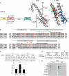
GGA GAT binding to PI4P. (A) GGA domain organization and structure. Left, the modular organization of the GGA domains. The GGA1 domain boundaries are as defined by Suer et al. (2003). The GAT domain is further divided into the N-GAT and C-GAT domains, which bind Arf1 and Ub, respectively. Our data showed that C-GAT also binds PI4P. Middle, a ribbon diagram of the X-ray crystal structure of GGA1 GAT, adapted from Collins et al. (2003). The GAT domain has four helices (named α1–4). The N-GAT domain has a hook-helix (α1 and part of α2), and the C-GAT domain has a three-helix bundle that is comprised of a portion of α2 and the entire α3 and α4. The PI4P-binding residues, GGA1 R260 and R265 (equivalent to GGA2 R276 and R281) are highlighted in red. We showed the GGA1 GAT structure instead of the GGA2 GAT structure here because only the former has been solved at the atomic level. Right, an expanded view of the GGA1 C-GAT structure. The C-GAT domain is rotated slightly in order to display the relation between the Ub-binding sites and PI4P-binding residues. Red, PI4P-binding residues; blue and green, Ub-binding residues in sites 1 and 2, as defined in yeast (Bilodeau et al., 2004). Additional site 1 residues identified by X-ray crystallography of the mammalian GGA GAT:Ub (Bilodeau et al., 2004; Kawasaki et al., 2005; Prag et al., 2005) that fall outside of these regions are not highlighted to simplify the presentation (see panel B). The site 1 residues were observed to bind Ub in the GGA3 GAT:Ub structure by Prag et al. (2005) and Kawasaki et al. (2005). The site 2 binding residues were identified by site-directed mutagenesis (Bilodeau et al., 2004; Puertollano and Bonifacino, 2004; Shiba et al., 2004). (B) Sequence alignment of the human and yeast GAT domains. Rectangles, helices in GAT; dotted lines, linkers between helices. The boundaries used to generate the N-GAT and C-GAT are indicated. Red letters denote basic amino acids that were conserved among different species. GGA1 R260 and R265 are conserved in mammalian and yeast GGAs, except that there is a two-residue offset in the yeast proteins. Red arrowheads, PI4P binding residues identified in this study. Blue lines, Ub-binding sites 1 and 2 that were identified in yeast (Bilodeau et al., 2004). Red lines, site 1 residues observed to bind Ub in GGA3 GAT:Ub crystals (Kawasaki et al., 2005; Prag et al., 2005). There is currently no crystal structure of GAT site 2 bound to Ub. The mammalian site 2 residues identified by site-directed mutagenesis (Bilodeau et al., 2004; Puertollano and Bonifacino, 2004; Shiba et al., 2004) are not highlighted here. (C) Comparing the binding of GGA2 domains to liposomes. 0.02 mg/ml purified GGA2 GST-VHS-GAT, GAT, and VHS were incubated with 0.2 mg/ml mixed lipid vesicles containing 15% PS or PI4P. The liposomes were collected by centrifugation and the bound GGA proteins were detected by Coomassie blue staining. The bottom panel shows the ratios of protein cosedimentated with PI4P versus PS liposomes (PI4P-binding index; mean ± SE, n = 5 or 6). (D) Comparing the binding of GGA1 and GGA2 GAT to PI4P-containing liposomes. GAT proteins (0.02 mg/ml) were incubated with increasing amount of liposomes. (E) Comparing the binding of GGA2 VHS and GAT domains to lipid dots. Proteins were incubated with PIP-arrays (Echelon, dotted with lipids ranging from 100 to 1.6 pmol), and the bound GGA domains were detected with anti-GST antibody.
Figure 3.
Figure 3.
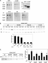
Identification of the GAT PI4P-binding site. (A) Recombinant GGA GST-GAT and its subdomains were incubated with nitrocellulose membranes dotted with 100, 50, and 25 pmol lipids. These lipid dot blots were made in the laboratory. A Coomassie blue–stained gel of the recombinant GST-GAT and GST-C-GAT used is shown. (B) Comparing the binding of wt and mutant GGA2 GAT to liposomes. 0.02 mg/ml (0.4 μM) GGA2 GST-GAT was incubated with 0.2 mg/ml mixed lipid vesicles. The vesicles were sedimented by centrifugation and bound proteins were detected by Coomassie blue staining after SDS-PAGE. The bottom panel shows the ratios of protein cosedimentated with PI4P versus PS liposomes (PI4P-binding index), as mean ± SE from two to four experiments. (C) Comparing the effect of Arf1-GTP on GAT binding to PS liposomes. PS liposomes were preloaded with or without recombinant myr-Arf1-GTPγS. The percent of GAT sedimented was shown in the right panel (mean ± SE, n = 3). **p = 0.003, *p = 0.02, NS (not significant), p = 0.15.
Figure 4.
Figure 4.
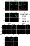
The requirements for GGA targeting to the TGN. (A) GAT targeting. Cells were transfected with myc-tagged GGA2 GAT wt or mutant cDNA. Arrows indicate cells overexpressing low amounts of GAT R281A. (B) Full-length GGA2 targeting. Cells were transfected with myc-tagged wt or mutant GGA2 cDNA. Left panels, immunofluorescence staining. Right panels, membrane fractionation, as described in Materials and Methods. Supernatant (S) and membrane pellets (P) were analyzed by Western blotting with myc and actin antibodies. (C) GGA1 RNAi rescue. Cells were initially transfected with control or GGA1 siRNA and then subsequently retransfected with either a myc-tagged empty vector (mock), the wt myc-GGA1 or myc-GGA1 R260A cDNA. GGA1 depletion was confirmed by Western blotting in C′. Actin was included as a loading control. The percentage of cells with the expanded TGN46 phenotype is indicated. Data were from a typical experiment and similar results were obtained in three independent experiments. (D) The GGA2 N-GAT and C-GAT domains were less enriched in the TGN than the GAT domain. Scale bar, 20 μm.
Figure 5.
Figure 5.
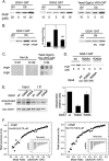
PI4P promotes GAT binding to Ub. (A) Ub-agarose binding. Mammalian GST-GAT or yeast His-VHS-GAT, 0.5 μM, was incubated with increasing amounts of PI4P in the presence of either protein A-agarose (Ctrl) or Ub-agarose. Bound proteins were detected with antibodies by Western blotting. (B) GAT binding to Ub-agarose was preferentially increased by PI4P. GST GGA1 GAT, 0.5 μM, was incubated with 20 μM PI4P or PI3P (left and middle panels) or with PC/PE liposomes containing either PS or PI4P (right panel). PI4P increased GGA1 GAT binding to Ub by 3.73 ± 0.8-fold and PI3P 1.5 ± 0.3-fold (n = 3). *p = 0.024. NS, p = 0.775. (C) His-Ub did not bind PI4P on lipid dot blots. His-Ub (at 8 or 40 nM) and His-VHS-GAT from yeast Gga1p (at 8 nM) were incubated with lipid dots made in the laboratory (lipids: 100, 50, and 25 pmol), and bound proteins were detected with anti-His antibody. (D) PI4P did not promote mutant GAT binding to Ub-agarose. GGA1 GAT wt, R265A, and R260A (0.25 μM) were incubated with Ub-agarose in the presence of 15 μM PI4P. The proteins used for the binding studies are shown (input). (E) GGA1 ubiquitination in vivo. HeLa cells were cotransfected with myc-tagged wt or mutant GGA1 and HA-Ub cDNAs. Myc-GGA1 was immunoprecipitated and blotted with anti-myc antibody to detect GGA1 and anti-HA to detect Ub associated with GGA1. The extent of ubiquitination for each sample was expressed as a ratio of the intensity of the HA-Ub to myc-GGA1 signals in the myc-GGA1 immunoprecipitates, and the value for the wt GGA1 is defined as 1.0. Data shown are mean ± SE (n = 3). (F) ITC analysis. GGA1 GST-GAT and bovine Ub were each preincubated with 50 μM water-soluble diC8PI4P before mixing. The kcal/mol of Ub as a function of the molar ratio of Ub to GGA1 GAT is shown, and the curves were fitted by using a one-site model. The Kd values shown are mean ± SE of five experiments using several different preparations of GGA1 GAT. The insets show the heat change elicited by successive injections of Ub into a chamber containing the GAT1 solution.

Similar articles

Cited by

References

    1. Akutsu M., Kawasaki M., Katoh Y., Shiba T., Yamaguchi Y., Kato R., Kato K., Nakayama K., Wakatsuki S. Structural basis for recognition of ubiquitinated cargo by Tom1-GAT domain. FEBS Lett. 2005;579:5385–5391. - PubMed
    1. Barylko B., Gerber S. H., Binns D. D., Grichine N., Khvotchev M., Sudhof T. C., Albanesi J. P. A novel family of phosphatidylinositol 4-kinases conserved from yeast to humans. J. Biol. Chem. 2001;276:7705–7708. - PubMed
    1. Behnia R., Munro S. Organelle identity and the signposts for membrane traffic. Nature. 2005;438:597–604. - PubMed
    1. Bilodeau P. S., Winistorfer S. C., Allaman M. M., Surendhran K., Kearney W. R., Robertson A. D., Piper R. C. The GAT domains of clathrin-associated GGA proteins have two ubiquitin binding motifs. J. Biol. Chem. 2004;279:54808–54816. - PMC - PubMed
    1. Bonifacino J. S. The GGA proteins: adaptors on the move. Nat. Rev. Mol. Cell Biol. 2004;5:23–32. - PubMed

Publication types

MeSH terms

LinkOut - more resources