. 2007 Jul;18(7):2687-97.
doi: 10.1091/mbc.e05-09-0873. Epub 2007 May 9.

Recycling endosomes of polarized epithelial cells actively sort apical and basolateral cargos into separate subdomains

Affiliations

Recycling endosomes of polarized epithelial cells actively sort apical and basolateral cargos into separate subdomains

Anthony Thompson et al. Mol Biol Cell. 2007 Jul.

Abstract

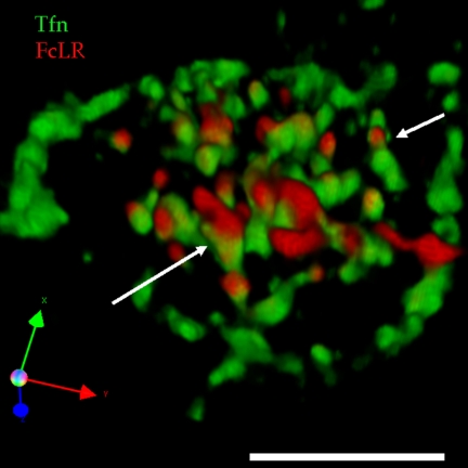
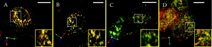
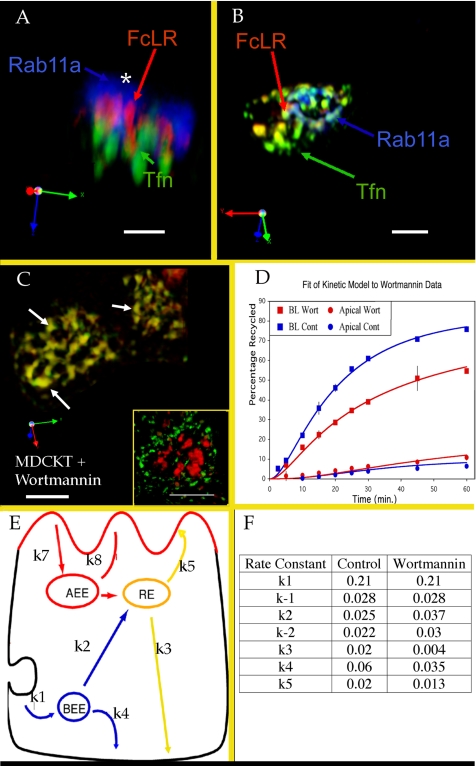
The plasma membranes of epithelial cells plasma membranes contain distinct apical and basolateral domains that are critical for their polarized functions. However, both domains are continuously internalized, with proteins and lipids from each intermixing in supranuclear recycling endosomes (REs). To maintain polarity, REs must faithfully recycle membrane proteins back to the correct plasma membrane domains. We examined sorting within REs and found that apical and basolateral proteins were laterally segregated into subdomains of individual REs. Subdomains were absent in unpolarized cells and developed along with polarization. Subdomains were formed by an active sorting process within REs, which precedes the formation of AP-1B-dependent basolateral transport vesicles. Both the formation of subdomains and the fidelity of basolateral trafficking were dependent on PI3 kinase activity. This suggests that subdomain and transport vesicle formation occur as separate sorting steps and that both processes may contribute to sorting fidelity.


Figures

Figure 1.
Figure 1.
Recycling Endosomes of polarized cells have subdomains. (A) Early endosomes (EE) and RE in MDCKT cells labeled with timed pulses/chases of basolaterally applied Tfn. Tfn (green) was internalized for 22.5 min to label REs, the cells were chilled, Alexa 546-Tfn (red) was bound to the basolateral surface, cells were rewarmed to 37°C for an additional 2.5 min to label EEs. (B) MDCKT cells infected with adeno FcLR(5-22) and induced with butyrate, then labeled with apical anti-FcLR(5-22) (red), and basolateral Tfn (green), and fixed with PFA. Cargoes segregate into RE subdomains. Serial confocal sections were reconstructed into this 3D projection. Inset is enlarged RE. (C) MDCK cells infected with adeno TfnR and adeno FcLR(5-22), labeled as in B but not induced with butyrate. (D) MDCKT cells infected induced and labeled as in B but imaged live (without fixation). Triple arrows indicate rotation of axes. Bars are 10 μm.
Figure 2.
Figure 2.
RE subdomains are a feature of polarized cells. (A) In nonpolarized CHOT and HeLa (inset) cell lines, apical and basolateral cargoes labeled as with Tfn (green) and anti-FcLR(5-22) (red) do not segregate into RE subdomains. Red areas represent EEs, which remain labeled by the antibody to FcLR(5-22). Arrow indicates area of overlap. (B) In a rat hippocampal neuron, apical and basolateral cargoes (labeled as in A) segregate into RE subdomains. Pulse period was 108 min at 37°C, chase was 25 min. Inset, enlarged RE. Triple arrows indicate rotation of axes. Bars are 10 μm.
Figure 3.
Figure 3.
Cargo in RE subdomains segregates by destination rather than by membrane of origin. MDCKT cells expressing FcLR(5-22) were labeled apically with anti FcLR(5-22) (red) and Tfn (blue), and basolaterally with Tfn (green). Label was chased for 25 min at 37°C. High levels of all labels make EEs visible in this protocol. (A) Apical FcLR(5-22) versus basolaterally bound Tfn (green). (B) Apically bound (red) versus basolaterally bound (green) Tfns. (C) Apically bound FcLR(5-22) (red) versus apically bound Tfn (green). (D) Combined projection of all three dyes. Triple colocalization is white. Arrowhead indicates a colocalization of Tfns with segregation of FcLR(5-22). Triple arrows indicate rotation of axes. Bars, 10 μm.
Figure 4.
Figure 4.
Transcytotic NgCAM colocalizes with FcLR(5-22), but not with Tfn, in REs. (A) NgCAM trafficking through the MDCKT cell. Nascent NgCAM is delivered from the Golgi to the basolateral surface where it is phosphorylated (purple circle), which inactivates the basolateral sorting determinant. Basolateral NgCAM is then endocytosed and delivered, as an apically destined protein, to the REs. From there, it is delivered to the apical surface where it remains. Once bound to NgCAM at the basolateral surface, antibody 8D9 remains with the protein during basolateral to apical transcytosis. (B) In hippocampal neuron REs, axonally targeted NgCAM (red) segregates from somatodendritic Tfn (green). Inset shows subdomains, with NgCAM and TFN segregated into different subdomains of the same REs (arrows). (C) In MDCK cell REs, basolaterally labeled NgCAM (blue) colocalizes with apically labeled FcLR(5-22) (red) but not with basolaterally labeled Tfn (green). Three cells are shown. NgCAM and FcLR largely colocalize (purple), but segregate from basolaterally bound Tfn (green) at arrows. Yellow box indicates area enlarged in D. (D) REs from left cell in C, shown in label pairs. Each pair has been rendered in red/green for better contrast. Arrow indicates a single RE that illustrates the segregation of apically destined NgCAM and FcLR(5-22) from basolaterally destined Tfn. Triple arrows indicate rotation of axes. Bars, 10 μm.
Figure 5.
Figure 5.
Slowing basolateral traffic through the REs does not abolish subdomain formation. MDCKT cells were labeled on ice with apical anti-FcLR(5-22) (red) and basolateral Tfn (green). The cells were treated on ice for 30 min with AlF4, and both labels were chased into the cells at 37°C in the presence of AlF4. A 3D reconstruction of a typical cell is shown. Arrows indicate continued presence of apical and basolateral subdomains within the same RE. Bar, 10 μm. Triple arrow is axis of rotation.
Figure 6.
Figure 6.
Formation of polarized RE subdomains. MDCKT cells expressing FcLR(5-22) are labeled basolaterally with Tfn (green) and apically with anti-FcLR(5-22). 3D reconstructions of typical cells are shown (A) 1 d, (B) 2 d, (C) 3 d, and (D) 4 d after plating. Insets are magnified views of boxed regions in each frame. Yellow is overlap of the two markers. Bars, 10 μm.
Figure 7.
Figure 7.
AP-1B is not required for subdomain formation. LLC-PK1 cells lacking the AP-1B adaptor component μ1-B, were stably transfected with μ1-A (A) or μ1-B (B). Cells were labeled apically with anti-FcLR(5-22) (red) and basolaterally with Tfn (green). Arrows indicate representative subdomains. Inset is enlarged RE. Pictures are 3D reconstructions of 30 confocal sections. Triple arrows indicate rotation of axes. Bars, 10 μm.
Figure 8.
Figure 8.
Subdomain formation is not Rab11a dependent but is wortmannin sensitive. (A) 3D reconstruction of a single typical MDCKT cell apically labeled with anti-FcLR(5-22) (red), basolaterally with Tfn (green) by immunofluorescence for Rab11a (blue). Asterisk indicates largely cytosolic pool of Rab11a. (B) 3D reconstruction of a typical triple-labeled cell as in A, but treated with saponin on ice before fixation, and with the cytosolic component of Rab11a (which does not overlap with either a basolateral or apical cargo marker) removed from the digital reconstruction. (C) wortmannin-treated REs in two MDCKT cells labeled with Tfn (green) and anti-FcLR(5-22) (red). White arrows indicate example REs. Inset shows 3D reconstruction of wortmannin treated MDCKT cell labeled with Tfn for 2.5 min (green) or 25 min (red). (D) Kinetic analysis of wortmannin effect on Tfn recycling. Control basolateral recycling (upper blue squares, line), control transcytosis (lower blue circles, line), basolateral recycling in the presence of wortmannin (upper red squares, line) and transcytosis in the presence of wortmannin (lower red circles, line). Data points represent an average of six experiments. Error bars, 1 SD. Lines are curves fit from the mathematical model to the data. (E) Cartoon of the mathematical model. Rate constants are shown. Note that k−1 represents Tfn returned to the surface without ever passing through an acidified endosome, whereas k4 represents Tfn that has passed through EEs and is released upon reaching the cell surface. (F) Values derived for each rate constant. Internalization rates (k1, k−1) were determined directly as surface clearance of acid-strippable counts from the basolateral surface (separate unpublished data, n = 3). Note changes in EE and RE to plasma membrane rates.

Similar articles

Cited by

References

    1. Anderson E., Maday S., Sfakianos J., Hull M., Winckler B., Sheff D., Folsch H., Mellman I. Transcytosis of NgCAM in epithelial cells reflects differential signal recognition on the endocytic and secretory pathways. J. Cell Biol. 2005;170:595–605. - PMC - PubMed
    1. Ang A. L., Taguchi T., Francis S., Folsch H., Murrells L. J., Pypaert M., Warren G., Mellman I. Recycling endosomes can serve as intermediates during transport from the Golgi to the plasma membrane of MDCK cells. J. Cell Biol. 2004;167:531–543. - PMC - PubMed
    1. Barbero P., Bittova L., Pfeffer S. R. Visualization of Rab9-mediated vesicle transport from endosomes to the trans-Golgi in living cells. J. Cell Biol. 2002;156:511–518. - PMC - PubMed
    1. Besterman J. M., Low R. B. Endocytosis: a review of mechanisms and plasma membrane dynamics. Biochem. J. 1983;210:1–13. - PMC - PubMed
    1. Bomsel M., Parton R., Kuznetsov S. A., Schroer T. A., Gruenberg J. Microtubule- and motor-dependent fusion in vitro between apical and basolateral endocytic vesicles from MDCK cells. Cell. 1990;62:719–731. - PubMed

Publication types

Substances

LinkOut - more resources