. 2007 May 22;104(21):8869-74.
doi: 10.1073/pnas.0703268104. Epub 2007 May 10.

The tumor suppressor CYLD regulates entry into mitosis

Affiliations

The tumor suppressor CYLD regulates entry into mitosis

Frank Stegmeier et al. Proc Natl Acad Sci U S A. .

Abstract

Mutations in the cylindromatosis (CYLD) gene cause benign tumors of skin appendages, referred to as cylindromas. The CYLD gene encodes a deubiquitinating enzyme that removes Lys-63-linked ubiquitin chains from I kappa B kinase signaling components and thereby inhibits NF-kappaB pathway activation. The dysregulation of NF-kappaB activity has been proposed to promote cell transformation in part by increasing apoptosis resistance, but it is not clear whether this is CYLD's only or predominant tumor-suppressing function. Here, we show that CYLD is also required for timely entry into mitosis. Consistent with a cell-cycle regulatory function, CYLD localizes to microtubules in interphase and the midbody during telophase, and its protein levels decrease as cells exit from mitosis. We identified the protein kinase Plk1 as a potential target of CYLD in the regulation of mitotic entry, based on their physical interaction and similar loss-of-function and overexpression phenotypes. Our findings raise the possibility that, as with other genes regulating tumorigenesis, CYLD has not only tumor-suppressing (apoptosis regulation) but also tumor-promoting activities (enhancer of mitotic entry). We propose that this additional function of CYLD could provide an explanation for the benign nature of most cylindroma lesions.


Conflict of interest statement

The authors declare no conflict of interest.

Figures

Fig. 1.
Fig. 1.
CYLD regulates premitotic cell-cycle progression independent of NF-κB pathway regulation. (A and B) HeLa cells were transfected with siRNAs (numbers refer to different oligos), treated with 100 nM Taxol 48 h posttransfection, and fixed for visual inspection 24 h after taxol addition. The percentage of nonmitotic cells (A) and cells with multilobed or interphase nuclei (B) was quantified. Mad2 siRNAs were used as a positive control for a spindle checkpoint regulator. The values represent averages of three independent experiments (n = 100, error bars ± 1 SD). The Western blot in B shows the extent of CYLD protein depletion 48 h after siRNA transfection. (C) Representative images of HeLa cells transfected with siRNAs targeting luciferase (control) or CYLD (oligo1) that illustrate the premitotic nuclear morphology of CYLD-depleted cells. (Magnification: ×20.) (D) Transfection of siRNA-resistant wild-type HA-tagged CYLD (CYLD-WT), but not the catalytically inactive-mutant (CYLD-ci), rescues the premitotic delay of CYLD siRNA-treated cells. HeLa cells were transfected with the indicated siRNAs (CYLD siRNA targets the 3′ UTR). After 24 h, the rescuing plasmids were cotransfected with H2B-GFP. Forty-eight hours after siRNA transfection, 100 nM taxol was added for an additional 24 h before fixation. GFP-positive cells were analyzed for the percentage of nonmitotic cells (n = 100, error bars ± 1 SD). The Western blot shows similar expression levels of wild-type and mutant HA-CYLD. (E) Down-regulation of NF-κB signaling does not rescue CYLD's cell-cycle defect. HeLa cells were transfected with the indicated siRNAs. Twenty-four hours later, the indicated NF-κB inhibitory plasmids were cotransfected with H2B-GFP. GFP-positive cells were analyzed for the percentage of nonmitotic cells (n = 100, error bars ± 1 SD). The Western blot shows expression of NF-κB inhibitory constructs.
Fig. 2.
Fig. 2.
CYLD is required for timely entry into mitosis. (A) HeLa cells were treated with siRNAs targeting luciferase (−) or CYLD (Cyl), synchronized in mitosis (100 ng/ml nocodazole), and released into fresh medium, and nocodazole was readded 6 h after release to arrest cells in the next mitosis. For detailed synchronization protocol see SI Fig. 6D. Cell extracts were collected at the indicated time points and immunoblotted for CYLD, GAPDH, Cdc25C (arrowhead indicates phosphorylation form), and P-H3 (serves as mitotic marker). (B–D) HeLa cells were treated with the indicated siRNAs, synchronized at the G1/S transition by a double thymidine block, and released from the second thymidine block into medium containing 100 ng/ml Nocodazole (added 6 h after release) to arrest cells in mitosis. (B) FACS sorting for P-H3-positive cells was used to determine the percentage of mitotic cells at the indicated time points. (C) Cell extracts were collected and immunoblotted for the indicated proteins (P-Cdk1, antibody recognizing Y15-phosphorylated Cdk1). (D) Schematic of synchronization protocol applied in B–E. (E) FACS profiles (DNA staining) of control and CYLD-depleted cells showing that DNA replication occurs with normal kinetics in cells with reduced CYLD function.
Fig. 3.
Fig. 3.
CYLD is degraded as cells exit from mitosis. (A) HeLa cells were released from a mitotic arrest (100 ng/ml Nocodazole), and cell lysates were blotted with the indicated antibodies. The faster migration of Cdc27 (1 h time point) indicates dephosphorylation and serves as a marker for exit from mitosis. (B) HeLa cells were synchronized in mitosis (100 ng/ml Nocodazole, Noc), 10 mM Cycloheximide (CHX) was added (to prevent de novo protein synthesis), and Nocodazole was either released from (Noc release) or cultured in the continued presence of nocodazole (Noc arrest). Cell lysates were analyzed by Western blotting.
Fig. 4.
Fig. 4.
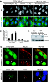
CYLD overexpression leads to accumulation of multinucleated cells. (A–C) U2OS TET-OFF cells stably expressing tetracycline-responsive wild-type (TET-CYLD-WT) or catalytically inactive CYLD mutant (TET-CYLD-ci) were either cultured in the presence (+DOX, repressed) or absence (−DOX, induced) of doxycycline (DOX, tetracycline analog). (A) Representative images taken 48 h after doxycycline treatment illustrate the presence of multinucleated nuclei after CYLD overexpression. (B) The percentage of cells with fragmented or multilobed nuclei was quantified (n > 100, error bars ± 1 SD). (C) A Western blot showing expression levels of wild-type and mutant CYLD. (D and E) CYLD localizes to the microtubule cytoskeleton in interphase and is enriched at the midbody (marked by arrow) during telophase. (D) HeLa cells were transiently transfected with GFP–CYLD and its localization was assessed by indirect immunofluorescence. Tubulin is stained in red. (E) HeLa cells stably expressing low levels of GFP-CYLD (green) were probed for tubulin (red) and stained for DNA (blue). (Magnifications: ×100.)
Fig. 5.
Fig. 5.
CYLD associates with Plk1 in vivo. (A) α-HA purification of stably integrated retrovirally expressed HA-Flag-CYLD from 293T cells shows that CYLD copurifies with multiple proteins not found in control purifications (HGS; see Materials and Methods). Ten percent of each HA peptide elution was analyzed by SDS/PAGE using a 4–12% gradient gel and silver stained. (B) Mass spectrometry analysis of purified CYLD-interacting proteins identifies Plk1. Total peptides identified in raw mass spectrometry data (with no filtering) are shown in red, and the five peptides found after stringent filtering (see Materials and Methods) are underlined. (C) Exogenously expressed CYLD associates with Plk1. HeLa cells were transiently transfected with the indicated MYC-tagged expression constructs and HA-Plk1. Anti-HA immunoprecipitates (IP) were probed with α-MYC (Upper) and α-HA antibodies (Lower). (D) Endogenous CYLD associates with Plk1. Plk1 was immunoprecipitated (IP) with α-Plk1 or α-CycB1 (control) antibodies, and the immunoprecipitates were probed for CYLD and Plk1.

References

    1. van Balkom ID, Hennekam RC. J Med Genet. 1994;31:321–324. - PMC - PubMed
    1. Bignell GR, Warren W, Seal S, Takahashi M, Rapley E, Barfoot R, Green H, Brown C, Biggs PJ, Lakhani SR, et al. Nat Genet. 2000;25:160–165. - PubMed
    1. Brummelkamp TR, Nijman SM, Dirac AM, Bernards R. Nature. 2003;424:797–801. - PubMed
    1. Kovalenko A, Chable-Bessia C, Cantarella G, Israel A, Wallach D, Courtois G. Nature. 2003;424:801–805. - PubMed
    1. Trompouki E, Hatzivassiliou E, Tsichritzis T, Farmer H, Ashworth A, Mosialos G. Nature. 2003;424:793–796. - PubMed

Publication types