. 2007 May 22;104(21):9018-23.
doi: 10.1073/pnas.0703229104. Epub 2007 May 10.

Angiotensin II up-regulates soluble epoxide hydrolase in vascular endothelium in vitro and in vivo

Affiliations

Angiotensin II up-regulates soluble epoxide hydrolase in vascular endothelium in vitro and in vivo

Ding Ai et al. Proc Natl Acad Sci U S A. .

Abstract

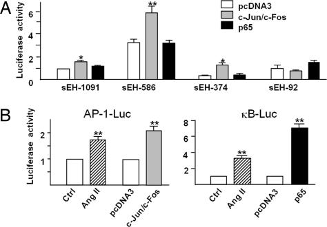
Epoxyeicosatrienoic acids (EETs), as metabolites of arachidonic acid, may function as antihypertensive and antiatherosclerotic mediators for vasculature. EETs are degraded by soluble epoxide hydrolase (sEH). Pharmacological inhibition and genetic ablation of sEH have been shown to increase the level of EETs, and treating angiotensin II (Ang II)-infused hypertension rats with sEH-selective inhibitors increased the levels of EETs, with attendant decrease in systolic blood pressure. To elucidate the mechanisms by which Ang II regulates sEH expression, we treated human umbilical vein endothelial cells (ECs) and bovine aortic ECs with Ang II and found increased sEH expression at both the mRNA and protein levels. Transient transfection assays showed that the activity of the human sEH promoter was increased in ECs in response to Ang II. Further analysis of the promoter region of the sEH gene demonstrated that treatment with Ang II, like overexpression of c-Jun/c-Fos, activates the sEH promoter through an AP-1-binding motif. The binding of c-Jun to the AP-1 site of the sEH promoter was confirmed by chromatin immunoprecipitation assays. In contrast, adenovirus overexpression of the dominant-negative mutant of c-Jun significantly attenuated the effects of Ang II on sEH induction. An elevated level of sEH was found in the aortic intima of both spontaneously hypertensive rats and Ang II-infused Wistar rats. Blocking Ang II binding to Ang II receptor 1 by losartan abolished the sEH induction. Thus, AP-1 activation is involved in the transcriptional up-regulation of sEH by Ang II in ECs, which may contribute to Ang II-induced hypertension.


Conflict of interest statement

Conflict of interest statement: B.D.H. founded Arête Therapeutics to develop sEH inhibitors.

Figures

Fig. 1.
Fig. 1.
Ang II induces sEH expression in ECs. (A and B) HUVECs (A) and BAECs (B) were treated with different concentrations of Ang II as indicated for 24 h or 100 nM Ang II for different periods of time. Cells were lysed, and proteins were resolved by 10% SDS/PAGE, transferred to a nitrocellulose membrane and probed with anti-sEH and anti-β-actin antibodies. Data are representative of three independent experiments. (C) HUVECs were incubated with 100 nM Ang II for 24 h. RNA was isolated and samples of total RNA analyzed by real-time RT-PCR with primers specific for human sEH, CYP2C9, or CYP2J2. β-Actin cDNA was used as an internal control. Data are means ± SD of the relative mRNA normalized to that of β-actin from three independent experiments.
Fig. 2.
Fig. 2.
Ang II activates the human sEH promoter in ECs. (A) Sequence of the 1.1-kb-cloned human sEH promoter and sketch of putative AP-1 and NFκB-binding sites. (B) BAECs were transfected with plasmids of sEH-Luc (−1091, −586, −374, and −92). (C) BAECs were transfected with sEH-586 or -586D. All transfected cells were then treated with 100 nM Ang II for 24 h. The β-gal plasmid was cotransfected as a transfection control. Promoter activities were measured by use of luciferase, which was normalized to β-gal. The results are expressed as the relative luciferase activities, of which activities of −1091-Luc in B and −586D-Luc in C are designed as 1. Data are mean ± SD of the relative luciferase activities from three independent experiments, each performed in triplicate (∗, P < 0.05).
Fig. 3.
Fig. 3.
AP-1 overexpression activates the human sEH promoter in ECs. (A) Plasmids of sEH-Luc (−1091, −586, −374, and −92) were cotransfected with expression plasmids of c-Jun, c-Fos, p65, or pcDNA3 in BAECs for 48 h. (B) BAECs were transfected with plasmids of 5 × NF-κB-Luc or 7 × AP-1-Luc for 24 h and then treated with 100 nM Ang II for 24 h. In parallel experiments, cells were cotransfected with the expression plasmids of c-Jun and c-Fos, p65, or pcDNA3 for 48 h. The β-gal plasmid was cotransfected in all experiments as a transfection control. Promoter activities measurement and data analysis were the same as those in Fig. 2.
Fig. 4.
Fig. 4.
AP-1 is involved in the Ang II-induced sEH in ECs. (A) Confluent HUVECs were infected with Ad-c-Jun for 24 h or treated with Ang II for 12 h. After cross-linking and sonication, nuclear proteins were extracted. ChIP assays involved use of anti-c-Jun antibody for IP, and normal rabbit IgG was used in control experiments. PCR involved use of sEH promoter-specific primers to detect binding of c-Jun to the sEH promoter. (B) HUVECs were infected with Ad-c-Jun at different multiplicity of infection for 48 h. (C) HUVECs were infected with Ad-TAM67 or Ad-TTA control virus for 24 h. Then, the cells were treated with 100 nM Ang II for another 24 h. Cell lysates were analyzed by Western blotting with use of anti-sEH, anti-c-Jun, or anti-β-actin antibodies. Results are representative of three independent experiments.
Fig. 5.
Fig. 5.
Level of sEH protein is elevated in the aortic intima of SHR rats. Wistar and SHR rats (180–280 g, male, n = 6) had free access to tap water supplemented with or without 2% NaCl for 14 days. (A) Systolic blood pressure was measured every 2 days until rats were killed. (B) Plasma Ang II levels were measured by use of an RIA kit. (C) Protein extracts of aorta intima from each rat were analyzed by Western blotting with anti-sEH and anti-β-actin antibodies in three individual animals from two separate sets of experiments. (D) The cross sections of the abdominal aorta from different treated rats were subjected to immunohistochemical staining with anti-sEH antibody. The results shown are representative of the rats from two separate sets of experiments. The sections were counterstained with hematoxylin.
Fig. 6.
Fig. 6.
Ang II infusion increases sEH protein in the aortic intima of Wistar rats. Wistar rats (180–280 g, male) received losartan (Los, 25 mg/kg per day) or PBS (Ctrl) by oral gavage for 6 days. A minipump was then implanted in the dorsal region to deliver either Ang II at 450 ng/kg per minute (Ang II) or PBS for 3 days. (A) Systolic blood pressure was measured daily after implantation. (B and C) At day 3, rats were killed, and protein extracts from intima (B) or media (C) of aortas were analyzed by Western blotting with anti-sEH and anti-β-actin antibodies. The density of each band was quantified with a densitometer, and the quantified data are mean ± SD of the relative mRNA normalized to that of β-actin in three individual animals from two separate sets of experiments.

References

    1. Spector AA, Fang X, Snyder GD, Weintraub NL. Prog Lipid Res. 2004;43:55–90. - PubMed
    1. Campbell WB, Gebremedhin D, Pratt PF, Harder DR. Circ Res. 1996;78:415–423. - PubMed
    1. Fisslthaler B, Popp R, Kiss L, Potente M, Harder DR, Fleming I, Busse R. Nature. 1999;401:493–497. - PubMed
    1. Hu S, Kim HS. Eur J Pharmacol. 1993;230:215–221. - PubMed
    1. Pokreisz P, Fleming I, Kiss L, Barbosa-Sicard E, Fisslthaler B, Falck JR, Hammock BD, Kim IH, Szelid Z, et al. Hypertension. 2006;47:762–770. - PMC - PubMed

Publication types

MeSH terms