

Neurally released pituitary adenylate cyclase-activating polypeptide enhances guinea pig intrinsic cardiac neurone excitability
- PMID: 17495034
- PMCID: PMC2075297
- DOI: 10.1113/jphysiol.2007.134965
Neurally released pituitary adenylate cyclase-activating polypeptide enhances guinea pig intrinsic cardiac neurone excitability
Abstract
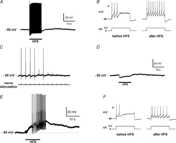
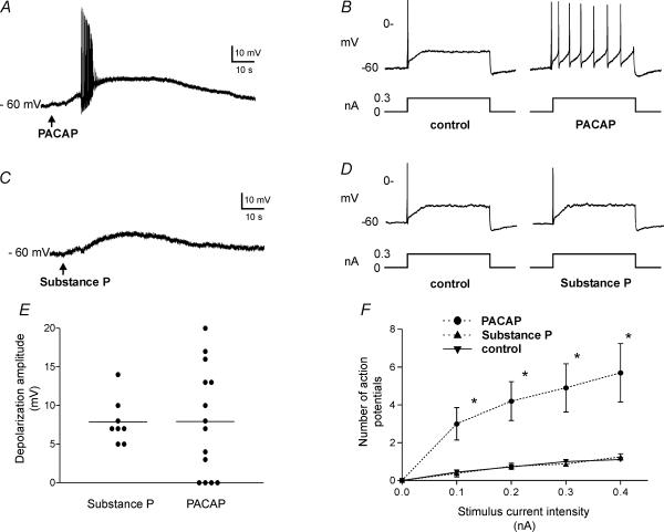
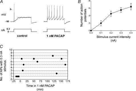
Intracellular recordings were made in vitro from guinea-pig cardiac ganglia to determine whether endogenous neuropeptides such as pituitary adenylate cyclase-activating polypeptide (PACAP) or substance P released during tetanic neural stimulation modulate cardiac neurone excitability and/or contribute to slow excitatory postsynaptic potentials (sEPSPs). When nicotinic and muscarinic receptors were blocked by hexamethonium and atropine, 20 Hz stimulation for 10 s initiated a sEPSP in all innervated neurones. In 40% of the cells, excitability was enhanced after termination of the sEPSP. This suggested that non-cholinergic receptor-mediated mechanisms contributed to the sEPSP and modulated neuronal excitability. Exogenous PACAP and substance P initiated a slow depolarization in the neurones whereas neuronal excitability was only increased by PACAP. When ganglia were treated with the PAC1 antagonist PACAP6-38 (500 nM), the sEPSP evoked by 20 Hz stimulation was reduced by approximately 50% and an enhanced excitability occurred in only 10% of the cells. These observations suggested that PACAP released from preganglionic nerve terminals during tetanic stimulation enhanced neuronal excitability and evoked sEPSPs. After addition of 1 nM PACAP to the bath, 7 of 9 neurones exhibited a tonic firing pattern whereas in untreated preparations, the neurons had a phasic firing pattern. PACAP6-38 (500 nM) diminished the increase in excitability caused by 1 nM PACAP so that only 4 of 13 neurones exhibited a tonic firing pattern and the other 9 cells retained a phasic firing pattern. These findings indicate that PACAP can be released by tetanic neural stimulation in vitro and increase the excitability of intrinsic cardiac neurones. We hypothesize that in vivo PACAP released during preganglionic firing may modulate neurotransmission within the intrinsic cardiac ganglia.
Figures
Similar articles
-
PAC1 Receptor Internalization and Endosomal MEK/ERK Activation Is Essential for PACAP-Mediated Neuronal Excitability.J Mol Neurosci. 2021 Aug;71(8):1536-1542. doi: 10.1007/s12031-021-01821-x. Epub 2021 Mar 6. J Mol Neurosci. 2021. PMID: 33675454 Free PMC article. Review.
-
Modulation of AMPA receptor-mediated ion current by pituitary adenylate cyclase-activating polypeptide (PACAP) in CA1 pyramidal neurons from rat hippocampus.Hippocampus. 2009 Jan;19(1):99-109. doi: 10.1002/hipo.20488. Hippocampus. 2009. PMID: 18727050
-
Pituitary adenylate cyclase-activating polypeptide expression and modulation of neuronal excitability in guinea pig cardiac ganglia.J Neurosci. 1998 Dec 1;18(23):9766-79. doi: 10.1523/JNEUROSCI.18-23-09766.1998. J Neurosci. 1998. PMID: 9822736 Free PMC article.
-
Pituitary adenylate cyclase activating polypeptide (PACAP) decreases neuronal somatostatin immunoreactivity in cultured guinea-pig parasympathetic cardiac ganglia.Neuroscience. 2004;126(2):335-46. doi: 10.1016/j.neuroscience.2004.04.007. Neuroscience. 2004. PMID: 15207351
-
PACAP-Induced PAC1 Receptor Internalization and Recruitment of Endosomal Signaling Regulate Cardiac Neuron Excitability.J Mol Neurosci. 2019 Jul;68(3):340-347. doi: 10.1007/s12031-018-1127-x. Epub 2018 Jul 27. J Mol Neurosci. 2019. PMID: 30054797 Free PMC article. Review.
Cited by
-
Is PACAP the major neurotransmitter for stress transduction at the adrenomedullary synapse?J Mol Neurosci. 2012 Oct;48(2):403-12. doi: 10.1007/s12031-012-9749-x. Epub 2012 May 18. J Mol Neurosci. 2012. PMID: 22610912 Free PMC article. Review.
-
PAC₁ receptors mediate positive chronotropic responses to PACAP-27 and VIP in isolated mouse atria.Eur J Pharmacol. 2013 Aug 5;713(1-3):25-30. doi: 10.1016/j.ejphar.2013.04.037. Epub 2013 May 9. Eur J Pharmacol. 2013. PMID: 23665113 Free PMC article.
-
Pretreatment with nonselective cationic channel inhibitors blunts the PACAP-induced increase in guinea pig cardiac neuron excitability.J Mol Neurosci. 2012 Nov;48(3):721-9. doi: 10.1007/s12031-012-9763-z. Epub 2012 Apr 14. J Mol Neurosci. 2012. PMID: 22528456 Free PMC article.
-
PAC1 Receptor Internalization and Endosomal MEK/ERK Activation Is Essential for PACAP-Mediated Neuronal Excitability.J Mol Neurosci. 2021 Aug;71(8):1536-1542. doi: 10.1007/s12031-021-01821-x. Epub 2021 Mar 6. J Mol Neurosci. 2021. PMID: 33675454 Free PMC article. Review.
-
Synaptic Plasticity in Cardiac Innervation and Its Potential Role in Atrial Fibrillation.Front Physiol. 2018 Mar 20;9:240. doi: 10.3389/fphys.2018.00240. eCollection 2018. Front Physiol. 2018. PMID: 29615932 Free PMC article. Review.
References
-
- Beker F, Weber M, Fink RHA, Adams DJ. Muscarinic and nicotinic ACh receptor activation differentially mobilize Ca2+ in rat intracardiac ganglion neurons. J Neurophysiol. 2003;90:1956–1964. - PubMed
-
- Brown D. M-currents: an update. Trends Neurosci. 1988;11:294–299. - PubMed
-
- Calupca MA, Vizzard MA, Parsons RL. Origin of pituitary adenylate cyclase-activating polypeptide (PACAP)-immunoreactive fibers innervating guinea pig parasympathetic cardiac ganglia. J Comp Neurol. 2000;423:26–39. - PubMed
Publication types
MeSH terms
Substances
Grants and funding
LinkOut - more resources
Full Text Sources