

Nano-scale dynamic recognition imaging on vascular endothelial cells
- PMID: 17496017
- PMCID: PMC1896235
- DOI: 10.1529/biophysj.107.109751
Nano-scale dynamic recognition imaging on vascular endothelial cells
Abstract
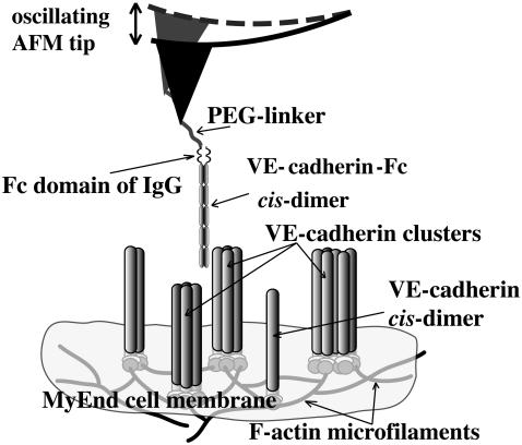
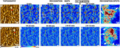
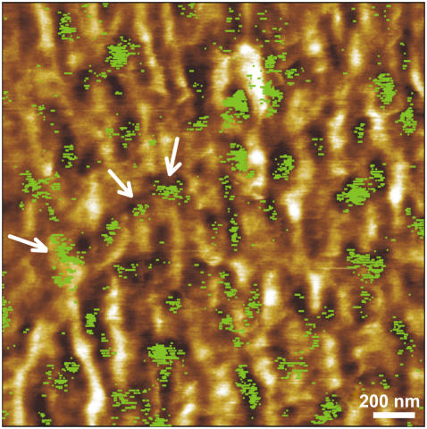
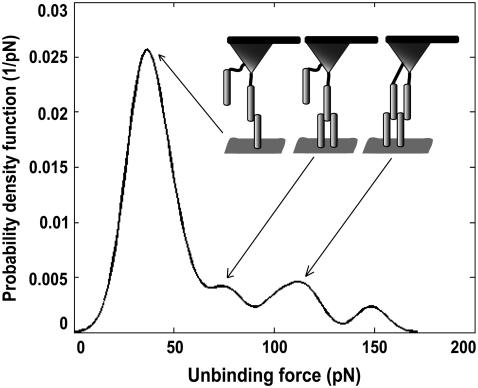
Combination of high-resolution atomic force microscope topography imaging with single molecule force spectroscopy provides a unique possibility for the detection of specific molecular recognition events. The identification and localization of specific receptor binding sites on complex heterogeneous biosurfaces such as cells and membranes are of particular interest in this context. Here simultaneous topography and recognition imaging (TREC) was applied to gently fixed microvascular endothelial cells from mouse myocardium (MyEnd) to identify binding sites of vascular endothelial (VE)-cadherin, known to play a crucial role in calcium-dependent, homophilic cell-to-cell adhesion. TREC images were acquired with magnetically oscillating atomic-force microscope tips functionalized with a recombinant VE-cadherin-Fc cis-dimer. The recognition images revealed single molecular binding sites and prominent, irregularly shaped dark spots (domains) with sizes ranging from 10 to 100 nm. These domains arose from a decrease of the oscillation amplitude during specific binding between active VE-cadherin cis-dimers. The VE-cadherin clusters were subsequently assigned to topography features. TREC represents an exquisite method to quickly obtain the local distribution of receptors on cellular surface with an unprecedented lateral resolution of 5 nm.
Figures
References
-
- Willing, K. I., S. O. Rizzoli, V. Westphal, R. Jahn, and S. W. Hell. 2006. STED microscopy reveals that synaptotagmin remains clustered after synaptic vesicle exocytosis. Nature. 440:935–939. - PubMed
-
- Koopman, M., A. Cambi, B. I. de Bakker, B. Joosten, C. G. Figdor, N. F. van Hulst, and M. F. Garcia-Parajo. 2004. Near-field scanning optical microscopy in liquid for high resolution single molecule detection on dendritic cells. FEBS Lett. 573:6–10. - PubMed
-
- Baumgartner, W., G. J. Schütz, J. Wiegand, N. Golenhofen, and D. Drenckhahn. 2003. Cadherin function probed by laser tweezer and single molecule fluorescence in vascular endothelial cells. J. Cell Sci. 116:1001–1011. - PubMed
Publication types
MeSH terms
Substances
LinkOut - more resources
Full Text Sources
Other Literature Sources
