Specific adsorption of osteopontin and synthetic polypeptides to calcium oxalate monohydrate crystals
- PMID: 17496021
- PMCID: PMC1948058
- DOI: 10.1529/biophysj.106.101881
Specific adsorption of osteopontin and synthetic polypeptides to calcium oxalate monohydrate crystals
Abstract
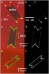
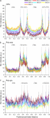
Protein-crystal interactions are known to be important in biomineralization. To study the physicochemical basis of such interactions, we have developed a technique that combines confocal microscopy of crystals with fluorescence imaging of proteins. In this study, osteopontin (OPN), a protein abundant in urine, was labeled with the fluorescent dye AlexaFluor-488 and added to crystals of calcium oxalate monohydrate (COM), the major constituent of kidney stones. In five to seven optical sections along the z axis, scanning confocal microscopy was used to visualize COM crystals and fluorescence imaging to map OPN adsorbed to the crystals. To quantify the relative adsorption to different crystal faces, fluorescence intensity was measured around the perimeter of the crystal in several sections. Using this method, it was shown that OPN adsorbs with high specificity to the edges between {100} and {121} faces of COM and much less so to {100}, {121}, or {010} faces. By contrast, poly-L-aspartic acid adsorbs preferentially to {121} faces, whereas poly-L-glutamic acid adsorbs to all faces approximately equally. Growth of COM in the presence of rat bone OPN results in dumbbell-shaped crystals. We hypothesize that the edge-specific adsorption of OPN may be responsible for the dumbbell morphology of COM crystals found in human urine.
Figures


Similar articles
-
Control of calcium oxalate crystal growth by face-specific adsorption of an osteopontin phosphopeptide.J Am Chem Soc. 2007 Dec 5;129(48):14946-51. doi: 10.1021/ja0745613. Epub 2007 Nov 10. J Am Chem Soc. 2007. PMID: 17994739
-
The osteopontin-controlled switching of calcium oxalate monohydrate morphologies in artificial urine provides insights into the formation of papillary kidney stones.Colloids Surf B Biointerfaces. 2016 Oct 1;146:296-306. doi: 10.1016/j.colsurfb.2016.06.030. Epub 2016 Jun 16. Colloids Surf B Biointerfaces. 2016. PMID: 27362921
-
Role of phosphate groups in inhibition of calcium oxalate crystal growth by osteopontin.Cells Tissues Organs. 2009;189(1-4):44-50. doi: 10.1159/000151430. Epub 2008 Aug 15. Cells Tissues Organs. 2009. PMID: 18703867
-
Molecular modulation of calcium oxalate crystallization.Am J Physiol Renal Physiol. 2006 Dec;291(6):F1123-31. doi: 10.1152/ajprenal.00136.2006. Am J Physiol Renal Physiol. 2006. PMID: 17082348 Review.
-
The role of macromolecules in the formation of kidney stones.Urolithiasis. 2017 Feb;45(1):57-74. doi: 10.1007/s00240-016-0948-8. Epub 2016 Dec 2. Urolithiasis. 2017. PMID: 27913854 Free PMC article. Review.
Cited by
-
Cementomimetics-constructing a cementum-like biomineralized microlayer via amelogenin-derived peptides.Int J Oral Sci. 2012 Jun;4(2):69-77. doi: 10.1038/ijos.2012.40. Epub 2012 Jun 29. Int J Oral Sci. 2012. PMID: 22743342 Free PMC article.
-
Peptides of Matrix Gla protein inhibit nucleation and growth of hydroxyapatite and calcium oxalate monohydrate crystals.PLoS One. 2013 Nov 12;8(11):e80344. doi: 10.1371/journal.pone.0080344. eCollection 2013. PLoS One. 2013. PMID: 24265810 Free PMC article.
-
Modulation of calcium oxalate dihydrate growth by selective crystal-face binding of phosphorylated osteopontin and polyaspartate peptide showing occlusion by sectoral (compositional) zoning.J Biol Chem. 2009 Aug 28;284(35):23491-501. doi: 10.1074/jbc.M109.021899. Epub 2009 Jul 6. J Biol Chem. 2009. PMID: 19581305 Free PMC article.
-
Face-specific binding of prothrombin fragment 1 and human serum albumin to inorganic and urinary calcium oxalate monohydrate crystals.BJU Int. 2009 Mar;103(6):826-35. doi: 10.1111/j.1464-410X.2008.08195.x. Epub 2008 Nov 13. BJU Int. 2009. PMID: 19021614 Free PMC article.
-
Morphogenesis and evolution mechanisms of bacterially-induced struvite.Sci Rep. 2021 Jan 8;11(1):170. doi: 10.1038/s41598-020-80718-y. Sci Rep. 2021. PMID: 33420384 Free PMC article.
References
-
- Lieske, J. C., and F. G. Toback. 2000. Renal cell-urinary crystal interactions. Curr. Opin. Nephrol. Hypertens. 9:349–355. - PubMed
-
- Shiraga, H., W. Min, W. J. VanDusen, M. D. Clayman, D. Miner, C. H. Terrell, J. R. Sherbotie, J. W. Foreman, C. Przysiecki, E. G. Nielson, and J. R. Hoyer. 1992. Inhibition of calcium oxalate crystal growth in vitro by uropontin: another member of the aspartic acid-rich protein superfamily. Proc. Natl. Acad. Sci. USA. 89:426–430. - PMC - PubMed
-
- Worcester, E. M., S. S. Blumenthal, A. M. Beshensky, and D. L. Lewand. 1992. The calcium oxalate crystal growth inhibitor protein produced by mouse kidney cortical cells in culture is osteopontin. J. Bone Miner. Res. 7:1029–1036. - PubMed
-
- Wesson, J. A., E. M. Worcester, J. H. Wiessner, N. S. Mandel, and J. G. Kleinman. 1998. Control of calcium oxalate crystal structure and cell adherence by urinary macromolecules. Kidney Int 53:952–957. - PubMed
-
- Wesson, J. A., V. Ganne, A. M. Beshensky, and J. G. Kleinman. 2005. Regulation by macromolecules of calcium oxalate crystal aggregation in stone formers. Urol. Res. 33:206–212. - PubMed
Publication types
MeSH terms
Substances
LinkOut - more resources
Full Text Sources
Other Literature Sources
Research Materials

