. 2007 Sep 15;93(6):2209-16.
doi: 10.1529/biophysj.106.102202. Epub 2007 May 11.

Gold-nanoparticle-assisted laser perturbation of chromatin assembly reveals unusual aspects of nuclear architecture within living cells

Affiliations

Gold-nanoparticle-assisted laser perturbation of chromatin assembly reveals unusual aspects of nuclear architecture within living cells

Aprotim Mazumder et al. Biophys J. .

Abstract

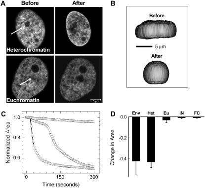
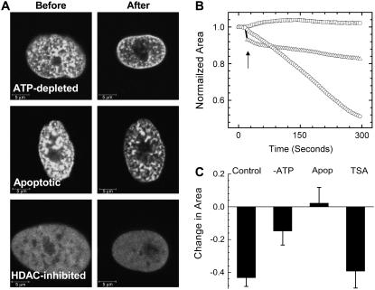
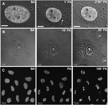
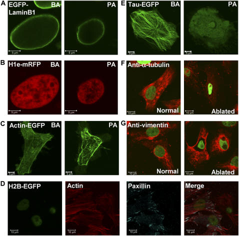
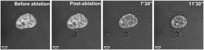
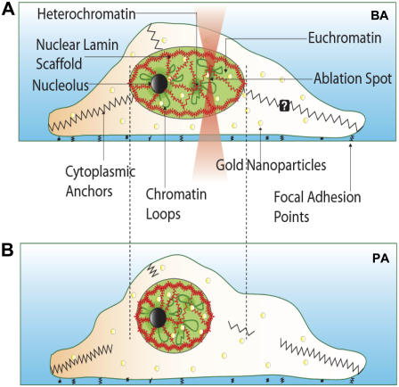
Chromatin organization within the nucleus is a vital regulator of genome function, yet its mechanical coupling to the nuclear architecture has remained elusive. To directly investigate this coupling, we locally modulated chromatin structure in living cells using nanoparticle-based laser perturbation. Unusual differences in the response of the cell nucleus were observed depending on the nuclear region that was perturbed--the heterochromatin, the euchromatin, and the nuclear envelope. This response varied under different conditions of cellular perturbations such as ATP depletion, apoptosis, and inhibition of histone deacetylases. Our studies implicate heterochromatin organization in imparting mechanical stability to the cell nucleus and suggest that nuclear size and shape are the result of interplay between nuclear and cytoplasmic anchors.


Figures

FIGURE 1
FIGURE 1
(A) Images before and after perturbation of heterochromatin and euchromatin in H2B-EGFP HeLa cells. The region indicated by the white arrows was exposed to ∼3 s of 835-nm NIR radiation, and the effect was monitored by imaging the H2B-EGFP fluorescence with the 488-nm line of an argon ion laser. The scale bar is equal to 5 μm. (B) Three-dimensional reconstructions of a cell made from a z-stack of confocal slices before and after perturbation shown from a primarily XZ perspective. Note that shrinkage in the XY plane is accompanied by a slight expansion in Z. (C) Representative plots of shrinkage dynamics for perturbation at the nuclear envelope (triangles), heterochromatin (circles), and euchromatin (squares) (D) Statistics of fractional change in area for perturbation at the nuclear envelope (Env), heterochromatin (Het), and euchromatin (Eu) (n = 12). Standard deviations are plotted as error bars. Also shown are similar statistics for isolated nuclei (IN) and fixed cells (FC) (n = 10), which did not show any shrinkage dynamics.
FIGURE 2
FIGURE 2
(A) Typical images before and after perturbation under different conditions of cellular perturbation: ATP depletion, staurosporine-induced apoptosis, and HDAC-inhibition by TSA. Scale bars are equal to 5 μm. (B) Representative plots of shrinkage dynamics under conditions of HDAC inhibition (circles), ATP depletion (triangles), and apoptosis (squares) on heterochromatin perturbation. A sharp decrease in area immediately on perturbation of ATP-depleted cells is indicated by the arrow. (C) Statistics of fractional change in area for perturbation under the different conditions indicated (n = 12), compared with shrinkage under conditions of heterochromatin perturbation in control cells (n = 12; from Fig. 1 D).
FIGURE 3
FIGURE 3
In all images, BA indicates “before perturbation”, and PA “postperturbation”. (A) Presence of cytoplasmic filaments (arrow) seen through time-lapse fluorescence-DIC images under conditions of heterochromatin perturbation. Scale bars are equal to 5 μm. (B) Time lapse DIC images to show that membrane blebs (arrows) appear at later time points on perturbation. (C) Chromatin fragmentation caused by subsequent apoptosis seen in fluorescence time-lapse images of a field of heterochromatin-perturbed cells. Scale bars are equal 20 μm. The time indicated in C is only approximate, as the cells were perturbed individually, and different cells are at different stages of progression toward cell death. Time points are indicated at the top of each picture.
FIGURE 4
FIGURE 4
State of the nuclear lamin on heterochromatin perturbation. HeLa cells were cotransfected with (A) EGFP-Lamin B1 and (B) H1e-mRFP. Images before and after perturbation are presented for the same cell. (C) Images of HeLa cells transiently transfected with Actin-EGFP before and after heterochromatin perturbation. Heterochromatin was identified by Hoechst staining of the DNA in these cases (images not shown). (D) H2B-EGFP-expressing HeLa cells were perturbed at the heterochromatin, fixed, and stained with an antibody against paxillin (visualized by a Cy5-labled secondary antibody) and filamentous actin bound by an Alexa-568–labeled phalloidin. Individual and merged images are presented. (E) Images of HeLa cells transiently transfected with Tau-EGFP as a microtubule marker before and after heterochromatin perturbation. As in C, heterochromatin was identified by Hoechst staining of the DNA. Images of H2B-EGFP expressing HeLa cells perturbed at the heterochromatin and stained with primary antibodies against (F) α-tubulin and (G) vimentin on separate plates. A Cy3-labled secondary antibody was used. Normal and perturbed cells were imaged on the same plate. Green indicates H2B-EGFP fluoresecnce, whereas red shows the respective cytoplasmic filaments. Scale bars are equal to 5 μm in A, B, C, and E. Scale bars are equal to 10 μm in D, F, and G.
FIGURE 5
FIGURE 5
Nuclear shrinkage and cytoplasmic blebs at late time points are also seen on cytoplasmic perturbations (scale bar = 5 μm). Fluorescence image of the nucleus of an H2B-EGFP HeLa cell is overlaid on a transmission DIC image. Time points are indicated at the top.
FIGURE 6
FIGURE 6
Model for the maintenance of cell nuclear architecture. (A) The nucleus is held under opposing tensions because of the chromatin and cytoplasmic filaments. On shining of an infrared laser on gold nanoparticle (yellow circles)-incorporated cells at the heterochromatin, there is a breaking of cytoplasmic contacts, resulting in a collapse of the nuclear volume depicted in B. The underlying lamin scaffold undergoes concomitant shrinkage but does not disintegrate during this process.

Similar articles

Cited by

References

    1. Cremer, T., and C. Cremer. 2001. Chromosome territories, nuclear architecture and gene regulation in mammalian cells. Nat. Rev. Genet. 2:292–301. - PubMed
    1. Foster, H. A., and J. M. Bridger. 2005. The genome and the nucleus: a marriage made by evolution. Genome organisation and nuclear architecture. Chromosoma. 114:212–229. - PubMed
    1. Horowitz-Scherer, R. A., and C. L. Woodcock. 2006. Organization of interphase chromatin. Chromosoma. 115:1–14. - PubMed
    1. Kosak, S. T., and M. Groudine. 2004. Gene order and dynamic domains. Science. 306:644–647. - PubMed
    1. Gerlich, D., J. Beaudouin, B. Kalbfuss, N. Daigle, R. Eils, and J. Ellenberg. 2003. Global chromosome positions are transmitted through mitosis in mammalian cells. Cell. 112:751–764. - PubMed

Publication types

LinkOut - more resources