. 2007 Sep 1;93(5):1557-70.
doi: 10.1529/biophysj.106.100248. Epub 2007 May 11.

Sodium channels: ionic model of slow inactivation and state-dependent drug binding

Affiliations

Sodium channels: ionic model of slow inactivation and state-dependent drug binding

Denis B Tikhonov et al. Biophys J. .

Abstract

Inactivation is a fundamental property of voltage-gated ion channels. Fast inactivation of Na(+) channels involves channel block by the III-IV cytoplasmic interdomain linker. The mechanisms of nonfast types of inactivation (intermediate, slow, and ultraslow) are unclear, although the ionic environment and P-loops rearrangement appear to be involved. In this study, we employed a TTX-based P-loop domain model of a sodium channel and the MCM method to investigate a possible role of P-loop rearrangement in the nonfast inactivation. Our modeling predicts that Na(+) ions can bind between neighboring domains in the outer-carboxylates ring EEDD, forming an ordered structure with interdomain contacts that stabilize the conducting conformation of the outer pore. In this model, the permeant ions can transit between the EEDD ring and the selectivity filter ring DEKA, retaining contacts with at least two carboxylates. In the absence of Na(+), the electrostatic repulsion between the EEDD carboxylates disrupts the permeable configuration. In this Na(+)-deficient model, the region between the EEDD and DEKA rings is inaccessible for Na(+) but is accessible for TMA. Taken together, these results suggest that Na(+)-saturated models are consistent with experimental characteristics of the open channels, whereas Na(+)-deficient models are consistent with experimentally defined properties of the slow-inactivated channels. Our calculations further predict that binding of LAs to the inner pore would depend on whether Na(+) occupies the DEKA ring. In the absence of Na(+) in the DEKA ring, the cationic group of lidocaine occurs in the focus of the pore helices' macrodipoles and would prevent occupation of the ring by Na(+). Loading the DEKA ring with Na(+) results in the electrostatic repulsion with lidocaine. Thus, there are antagonistic relations between a cationic ligand bound in the inner pore and Na(+) in the DEKA ring.


Figures
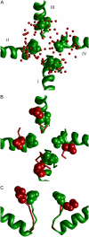
FIGURE 1
FIGURE 1
In the absence of TTX, the TTX receptor structure is unstable. (A) The extracellular view at the ensemble of the 26 lowest energy structures, which were obtained in a multi-MCM search started from the TTX receptor model. Red dots represent carbon atoms in the EEDD ring. For comparison, the outer carboxylates in the TTX receptor model are shown in the green-filled space. Roman numerals designate domains. (B and C) Top and side views of the superimposition of the TTX receptor model (green) with the lowest energy structure from the ensemble A (red). In the absence of TTX, the outer carboxylates repel each other and turn away from the pore axis.
FIGURE 2
FIGURE 2
Distance distribution from the pore axis of Na+ ions and carboxylate carbons in the EEDD ring. The distributions are calculated for the ion-saturated and ion-deficient models. In the Na+-saturated model, Na+ ions and outer carboxylate carbons most frequently appear 6–8 Å from the pore axis, which corresponds to the TTX receptor model. In the Na+-deficient model, carboxylate carbons move away from the pore axis and show a maximum at 10 Å from the pore axis, but a significant population remains at ∼7 Å.
FIGURE 3
FIGURE 3
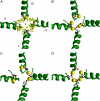
Stabilization of the EEDD ring by four Na+ ions. (A) Superposition of the 30 lowest energy complexes obtained by multi-MCM search. Na+ ions are shown as yellow dots. White-space-filled atoms are α-carbons of the outer carboxylates. Na+ ions are clustered between the carboxylates, whereas the pore lumen is poorly populated. (B and C) Representative examples from A that show yellow-space-filled Na+ ions interacting with neighboring carboxylates (sticks). (D) A possible conformation of the outer pore in which the outer carboxylates and four Na+ ions form a 16-membered cycle.
FIGURE 4
FIGURE 4
Na+ ions and EEDD and DEKA rings in the Na+-saturated (A) and Na+-excessive (B–D) models of the outer pore. A, In the Na+-saturated model, four Na+ ions form a 16-membered cycle with the outer carboxylates, and another Na+ is chelated between AspIp50 and GluIIp50 of the DEKA ring. Because of a large distance between the EEDD and DEKA rings, a single-step transition of Na+ between the rings is unlikely. (B) Insertion of an additional Na+ ion significantly perturbs coordination of Na+ ions. The 16-membered cycle at EEDD is broken and one of the Na+ ions is coordinated by two glutamates from the EEDD and DEKA rings. The model represents a possible transition state upon Na+ permeation via the outer pore. (C) Superposition of Na+ ions and the ammonium group of the DEKA Lys from 200 lowest energy structures collected in the multi-MCM search. For comparison, positions of seven K+ ions in the KcsA structure crystallized at high [K+] (11) are shown as red dots. (D) Distribution of Na+ ions and the ammonium group of the DEKA Lys along the outer pore. Vertical lines with numbers mark the outer-pore levels that correspond to K+-binding positions in KcsA (C). The average energy of Na+ ions and its standard deviations are shown.
FIGURE 5
FIGURE 5
Profiles of Na+ and TMA energy in the Na+-deficient model of the outer pore. The energy values are means from 12 independent calculations, with standard deviation shown as error bars. Vertical lines show K+ positions in KcsA (Fig. 4 C). Both Na+ and TMA experience unfavorable dehydration that increases as the ions move deeper into the outer pore (D). The electrostatic energy of Na+ has two minima at levels EEDD and DEKA (B). The weaker electrostatic energy between rings EEDD and DEKA is due to the inability of the outer carboxylates, which turn away from the pore axis, to escort Na+ toward the DEKA ring. It does not compensate the dehydration, causing an energy barrier for Na+. Unlike small Na+, a bulky TMA has favorable van der Waals interactions between the EEDD and DEKA rings that compensate the dehydration cost (C). The smooth profile of the total energy (A) evidences that TMA and compounds of similar size can reach as deep as the DEKA ring. Both Na+ and TMA profiles become highly rugged close to the DEKA ring due to competition with the resident Na+ ion there.
FIGURE 6
FIGURE 6
Paired Cys mutants of the TTX receptor model. The α-carbon tracing in the selectivity filter region is shown as red rods for the original TTX receptor model (35) and as green rods for MC-minimized models of the mutants. Side chains of Cys residues are space filled. (A and B) Extracellular views; (C and D) side views in which one repeat is not shown for clarity. (A and C) The model of the EIp53C/WIVp52C double mutant that minimally deviates from the TTX receptor model. (B and D) The model of the EIp53C/KIIIp50C double mutant that maximally deviates from the TTX receptor model.
FIGURE 7
FIGURE 7
Deformations of the TTX receptor model caused by Na+ and disulfide cross-linking. Pairs of residues cross-linked in the paired Cys mutants are indicated by their labels (see Table 1). (A) Disulfide cross-links cause up to 2 Å Cα-RMSD of the ascending limbs (positions p49–p54) versus the TTX receptor model as indicated by open bars. When compared to the TTX receptor model, distances between indicated Cα atoms usually decrease in the Na+-excessive model of the outer pore (solid bars) but increase in the Na+-deficient model (shaded bars). These predictions are consistent with the data of Xiong et al. (18) that the channel opening generally accelerates formation of disulfide cross-links, whereas slow inactivation prevents formation of certain S-S bonds. (B) Correlation between the distance change indicated by black bars at A and increase of the cross-linking rate upon the channel opening (18). For those paired Cys mutants, which deviate from the TTX receptor model up to 1 Å, the acceleration of the cross-linking reaction rate is small. For three paired Cys mutants, a big decrease of the Cα-Cα distances in the Na+-excessive model is observed. These mutants demonstrate significant acceleration of the cross-linking reaction upon the channel opening.
FIGURE 8
FIGURE 8
Binding of lidocaine in the Na+-deficient (A, C, and E) and Na+-saturated (B, D, and F) models of the inner pore. (A–D) Side views; (E and F) intracellular views. (A and B) Superposition of the 10 lowest energy structures obtained from 5,000 MCM trajectories. Lidocaine molecules are shown in a wire-frame mode with the protonated nitrogen as a red dot. (A) In the prevailing binding modes in the Na+-deficient model, the ligand's nitrogen is oriented toward the DEKA ring and occurs in the focus of macrodipoles from the pore helices. (B) Due to the electrostatic repulsion from the Na+ ion in the DEKA ring, the prevailing binding modes in the Na+-saturated model show the ligand's nitrogen oriented toward the cytoplasm. (C–F) Representative structures from A and B with a space-filled lidocaine molecule. Residues contributing to the interaction energy are shown as sticks. In the Na+-deficient model, the ligand binds tightly to IVS6 and IIIS6 and does not interact with IIS6 (C and E). In the Na+-saturated model, the ligand interacts with all four domains (D and F).
FIGURE 9
FIGURE 9
Energy profiles of lidocaine in the Na+-deficient and Na+-saturated models of the inner pore. For each model, two profiles were calculated with the ligand's ammonium group oriented either toward the DEKA ring (N-up profiles) or against the ring (N-down profiles). The energy values are means from 12 independent calculations with standard deviation shown as error bars. The vertical line marks K+ position 5 in the focus of macrodipoles of KcsA (Fig. 4 C). The ligand-receptor energy (A) has the lowest minimum in the Na+-deficient model with N-up orientation of lidocaine. This is due to electrostatic interactions (B), which are optimal when the ligand ammonium group occurs in the focus of macrodipoles from the pore helices in the absence of Na+ in the DEKA ring. This result agrees with the random search data (Fig. 8 A). Profiles of van der Waals (C) and solvation (D) energy components are rather similar. The increase of van der Waals energy close to the DEKA ring is due to the clash between the ligand and P-loops. The clash also results in significant differences between individual runs, which can be seen as large error bars.

Similar articles

Cited by

References

    1. Ulbricht, W. 2005. Sodium channel inactivation: molecular determinants and modulation. Physiol. Rev. 85:1271–1301. - PubMed
    1. Hoshi, T., W. N. Zagotta, and R. W. Aldrich. 1990. Biophysical and molecular mechanisms of Shaker potassium channel inactivation. Science. 250:506–507. - PubMed
    1. Vassilev, P. M., T. Scheuer, and W. A. Catterall. 1988. Identification of an intracellular peptide segment involved in sodium channel inactivation. Science. 241:1658–1661. - PubMed
    1. Kellenberger, S., J. W. West, T. Scheuer, and W. A. Catterall. 1997. Molecular analysis of the putative inactivation particle in the inactivation gate of brain type IIA Na+ channels. J. Gen. Physiol. 109:589–605. - PMC - PubMed
    1. Patton, D. E., J. W. West, W. A. Catterall, and A. L. Goldin. 1992. Amino acid residues required for fast sodium channel inactivation. Charge neutralizations and deletions in the III–IV linker. Proc. Natl. Acad. Sci. USA. 89:10905–10909. - PMC - PubMed

Publication types

LinkOut - more resources