. 2007 Sep 1;93(5):1719-35.
doi: 10.1529/biophysj.107.105528. Epub 2007 May 11.

Secondary and tertiary structure elasticity of titin Z1Z2 and a titin chain model

Affiliations

Secondary and tertiary structure elasticity of titin Z1Z2 and a titin chain model

Eric H Lee et al. Biophys J. .

Abstract

The giant protein titin, which is responsible for passive elasticity in muscle fibers, is built from approximately 300 regular immunoglobulin-like (Ig) domains and FN-III repeats. While the soft elasticity derived from its entropic regions, as well as the stiff mechanical resistance derived from the unfolding of the secondary structure elements of Ig- and FN-III domains have been studied extensively, less is known about the mechanical elasticity stemming from the orientation of neighboring domains relative to each other. Here we address the dynamics and energetics of interdomain arrangement of two adjacent Ig-domains of titin, Z1, and Z2, using molecular dynamics (MD) simulations. The simulations reveal conformational flexibility, due to the domain-domain geometry, that lends an intermediate force elasticity to titin. We employ adaptive biasing force MD simulations to calculate the energy required to bend the Z1Z2 tandem open to identify energetically feasible interdomain arrangements of the Z1 and Z2 domains. The finding is cast into a stochastic model for Z1Z2 interdomain elasticity that is generalized to a multiple domain chain replicating many Z1Z2-like units and representing a long titin segment. The elastic properties of this chain suggest that titin derives so-called tertiary structure elasticity from bending and twisting of its domains. Finally, we employ steered molecular dynamics simulations to stretch individual Z1 and Z2 domains and characterize the so-called secondary structure elasticity of the two domains. Our study suggests that titin's overall elastic response at weak force stems from a soft entropic spring behavior (not described here), from tertiary structure elasticity with an elastic spring constant of approximately 0.001-1 pN/A and, at strong forces, from secondary structure elasticity.


Figures

FIGURE 1
FIGURE 1
Schematic of the titin spring in muscle, and detailed view of titin's Z1Z2 domains. Shown is an electron micrograph image (courtesy R. Craig, University of Massachusetts) of the human muscle sarcomere and the schematic representation of titin along the sarcomere length. Titin is anchored with one of its ends (N-terminal) through the protein telethonin to the sarcomeric Z-disc and with its other end (C-terminal) to the M-line. Components of titin shown in this figure include the proximal Z1Z2 domains (circled), entropic PEVK region, the well-studied I27 (now numbered I91) domain in the I-band, and the Ig-Fn-III triad A168-170 of the A-band.
FIGURE 2
FIGURE 2
Two regimes of titin elasticity—soft and stiff nonlinear. Shown schematically is how titin responds to stretching forces of various magnitudes (,–29,31,32,34). At weak forces (<50 pN), the primary contribution of titin's response arises from the PEVK domains and the straightening of a bent and twisted titin chain. At high forces (>50 pN) titin exhibits a highly nonlinear elastic response. In this study we will relate the non-PEVK soft elasticity of titin to the so-called tertiary structure elasticity and the nonlinear stiff elasticity to the secondary structure elasticity. These two types of elasticities are defined in the text.
FIGURE 3
FIGURE 3
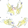
Experimentally derived conformers for titin Z1Z2 and a possible mode of tertiary structure elasticity. (A,B) Crystal structure (PDB code 2A38) for the N-terminal region of titin comprised of the tandem Z1 (purple) and Z2 (orange) domains in two crystallographically constrained conformations, termed closed and open, respectively. (C) NMR-RDC models of the experimentally observed semi-extended conformation for Z1Z2. The colors correspond to the NMR lowest (blue) and second lowest (red) energy conformers and the small angle x-ray scattering best-fit conformer (green) (58). (D) Highly schematic view of the tertiary structure elasticity of titin due to bending adjacent protein domains open and closed. In actual titin, not every linker may contribute high flexibility; some linkers might be short and stiff.
FIGURE 4
FIGURE 4
Stochastic modeling of titin's tertiary structure elasticity. (A) Schematic representation of a two-state multidomain chain. (B) Plot of the time-dependent end-to-end distribution given by Eq. 14, for N = 100, p = 0.3, b = 1, τR = 1, α0 = 45°, and X0 = 1.2 l0, which corresponds to an equilibrium length l0 ≈ 81 and initial length X0 ≈ 98. The initially extended chain relaxes back to its equilibrium length after a time comparable to τR. (C) Plot of the quadratic potential (Eq. 7) for the same system parameters and temperature T = 300 K.
FIGURE 5
FIGURE 5
Equilibrium simulation of Z1Z2 and convergence to NMR conformers. (A) The simulation system for the open form of crystallographically resolved Z1Z2 (Z1 in green; Z2 in orange; water is blue). (B) RMSD plot for simA2 comparing the deviation of secondary structural elements from the three experimentally derived structures (iiiv; see text) for Z1Z2 shown in Fig. 3. The colors, corresponding to those also used in Fig. 3 C, are selected as follows: the comparison of i, the simulated system, with ii is shown in blue, with iii in red, and with iv in green. The closest convergence of the MD model, i, to the experimental structures iiiv, arises between 25 ns and 38 ns. (C) Snapshots of the simulated Z1Z2 segment (simulation simA2) overlaid with iii at 0 ns, at ∼11.5 ns, and at ∼27 ns. A movie showing the entire trajectory for simA2 can be found in Supplementary Material.
FIGURE 6
FIGURE 6
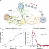
Adaptive biasing force (ABF) simulations of Z1Z2. The ABF simulations of titin Z1Z2 are depicted schematically (A); in the simulations, the crystallographic structure for the closed form of Z1Z2 (green) is hinged toward the open conformation and the potential of mean force (PMF) profile for this motion is calculated. The ABF simulations step through the states iviii. SimC1 determines the PMF between states i and ii, corresponding to an extension of 45–50 Å. Likewise, simC2 determines the PMF between states ii and iii with extension 50–55 Å, simC3 between states iii and iv with extension 55–60 Å, simC4 between states iv and v with extension 60–65 Å, simC5 between states v and vi with extension 65–75 Å, simC6 between states vi and vii with extension 75–80 Å and, finally, simC7 between vii and viii with extension 80–85 Å. The PMF profile over the extension range 45–85 Å, i.e., for transitioning between the closed and open state, is plotted (B), with I and II denoting intermediate states along the trajectory describing the stretched Z1Z2. The trough at II corresponds to the semi-extended conformer for Z1Z2 observed both in experiment and simulation (simA2). The distribution formula image defined through Eq. 33, is shown (C, red stars) along with the Gaussian distribution formula image defined in Eq. 34 (blue line). The dashed line at 45 Å denotes the beginning of the simulation range.
FIGURE 7
FIGURE 7
Charge-charge interactions within the titin Z1Z2 Ig-tandem. (A) The metal binding site for cadmium between Z1 and Z2 in the closed crystallographic conformer. The coordination between cadmium and Glu26, His128 of Z1, and Glu155 of Z2, stabilizes the constrained compact arrangement of tandem Ig-domains. Numerous weak interactions such as a Lys98:Glu100 salt bridge at the linker, captured during simA2 and shown (B), stabilize further the equilibrium conformation of Z1Z2.
FIGURE 8
FIGURE 8
Stretching individual Ig-domains of Z1 and Z2. (AC) Snapshots of the unfolding trajectory for titin Z1 under 350 pN constant force: (A) at t = 0 ns; (B) at t = 23.1 ns, i.e., the moment of simultaneous detachment of A and A′ strands (denoted by red and blue circles corresponding also to the strand color); and (C) at t = 23.5 ns, i.e., when the N-terminal region of Z1 extends. (D,E) Snapshots of the unfolding trajectory for titin Z2 under 350 pN constant force: (D) at t = 0 ns; (E) at t ∼ 19.9 ns when strand A detaches (red circle); and (F) at t ∼ 22.3 ns when strand A′ detaches (blue circle). An unfolding intermediate, in which the A′G strand holds briefly for ∼3 ns (see also Fig. 9 B) after the AB strand detaches, is seen during this unraveling of Z2, but not during the unraveling of Z1. After steps C and F, both Z1 and Z2 unfold quickly with little resistance, toward the elongated form shown in panel G.
FIGURE 9
FIGURE 9
Time evolution of the end-to-end distances of stretched Z1 and Z2. (A) Shown are the end-to-end distances of Z1 and Z2 for simulations simD1, simD2, simD3, simE1, simE2, and simE3. The traces demonstrate that unfolding occurs in a rupturelike event, i.e., rather suddenly, with a time delay after the onset of stretching that is longer for weaker forces. In particular, unfolding of Z1 and Z2 with a stretching force of 500 pN requires ∼7 ns simulation time, and with a stretching force of 350 pN requires ∼15 ns in simE2 and ∼30 ns in simD1-2 as well as simE1. Not shown here are the end-to-end distance traces from simD3 and simE3, in which Z1 and Z2 did not start to unfold within the 30-ns simulation period. (B) Shown is a detailed view of the unfolding event in simD1 (black) for Z1 and in simE1 (green) for Z2. The stepwise unraveling representative of an Ig unfolding intermediate (region circled) for Z2 (see also Fig. 8, D and E) reveals characteristic force plateaus corresponding to terminal β-strand rupture also seen for titin I91. Movies for the unfolding of Z1 and Z2 in simD2 and simE2, respectively, can be found in Supplementary Material.

Similar articles

Cited by

References

    1. Wang, K. 1996. Titin/connectin and nebulin: giant protein ruler of muscle structure and function. Adv. Biophys. 33:123–134. - PubMed
    1. Erickson, H. 1997. Stretching single protein modules: titin is a weird spring. Science. 276:1090–1093. - PubMed
    1. Maruyama, K. 1997. Connectin/titin, a giant elastic protein of muscle. FASEB J. 11:341–345. - PubMed
    1. Linke, W. 2000. Stretching molecular springs: elasticity of titin filaments in vertebrate striated muscle. Histol. Histopath. 15:799–811. - PubMed
    1. Tskhovrebova, L., and J. Trinick. 2003. Titin: properties and family relationships. Nat. Rev. Mol. Cell Biol. 4:679–689. - PubMed

Publication types

LinkOut - more resources