. 2007 Jul;189(14):5153-60.
doi: 10.1128/JB.00061-07. Epub 2007 May 11.

Temperature-hypersensitive sites of the flagellar switch component FliG in Salmonella enterica serovar typhimurium

Affiliations

Temperature-hypersensitive sites of the flagellar switch component FliG in Salmonella enterica serovar typhimurium

Takuji Mashimo et al. J Bacteriol. 2007 Jul.

Abstract

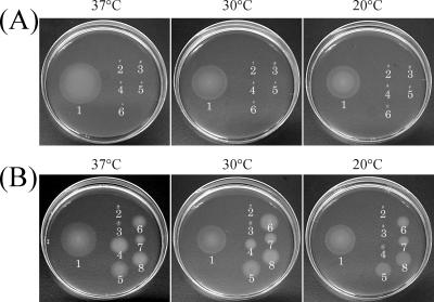
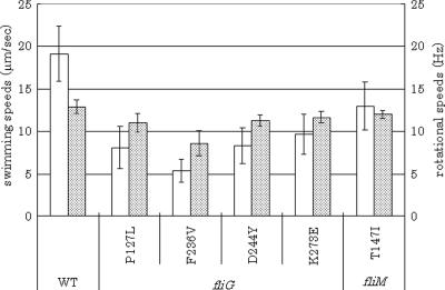
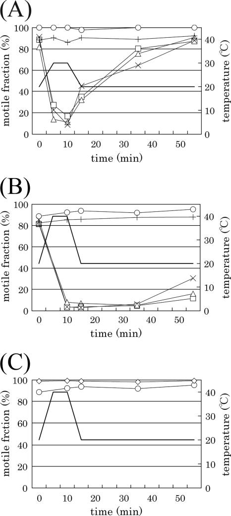
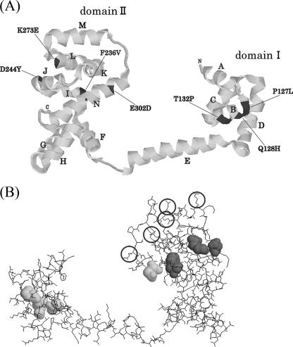
Three flagellar proteins, FliG, FliM, and FliN (FliGMN), are the components of the C ring of the flagellar motor. The genes encoding these proteins are multifunctional; they show three different phenotypes (Fla(-), Mot(-), and Che(-)), depending on the sites and types of mutations. Some of the Mot(-) mutants previously characterized are found to be motile. Reexamination of all Mot(-) mutants in fliGMN genes so far studied revealed that many of them are actually temperature sensitive (TS); that is, they are motile at 20 degrees C but nonmotile at 37 degrees C. There were two types of TS mutants: one caused a loss of function that was not reversed by a return to the permissive temperature (rigid TS), and the other caused a loss that was reversed (hyper-TS). The rigid TS mutants showed an all-or-none phenotype; that is, once a structure was formed, the structure and function were stable against temperature shifts. All of fliM and fliN and most of the fliG TS mutants belong to this group. On the other hand, the hyper-TS mutants (three of the fliG mutants) showed a temporal swimming/stop phenotype, responding to temporal temperature shifts when the structure was formed at a permissive temperature. Those hyper-TS mutation sites are localized in the C-terminal domain of the FliG molecules at sites that are different from the previously proposed functional sites. We discuss a role for this new region of FliG in the torque generation of the flagellar motor.


Figures

FIG. 1.
FIG. 1.
Swarm plates of TS switch (Mot) mutants. Strains were inoculated on 0.3% agar plates and incubated at 37°C for 9 h, at 30°C for 15 h, and at 20°C for 20 h. (A) Swarm plates of TS switch (Mot) mutants. 1, wild-type SJW1103; 2, SJW1774 [fliG(F236V)]; 3, SJW2827 [fliG(P127L)]; 4, SJW1770 [fliM(T147I)]; 5, SJW1771 [fliM(G132D)]; 6, SJW1799 [fliN(G103V)]. (B) Swarm plates of slow-swimming motAB mutants. 1, wild-type SJW1103; 2, MY6029 [motA(E33K)]; 3, MY6056 [motA(R90L)]; 4, MY6089 [motA(G176S)]; 5, MY6097 [motA(V207S)]; 6, MY6003 [motB(A37V)]; 7, MY6018 [motB(K28R)]; 8, MY6051 [motB(I29T)].
FIG. 2.
FIG. 2.
Swimming speeds and rotational speeds of group A TS mutants at 20°C. Empty columns show swimming speeds, and shaded columns show rotational speeds of wild-type SJW1103 (on the most left), fliG (Mot) mutants (P127L, F236V, D244Y, and K273E), and a fliM (Mot) mutant (T147I).
FIG. 3.
FIG. 3.
Swimming behaviors of TS mutants responding to temporal temperature shifts. Cells were grown at 20°C, shifted to the incubator at 30°C or 40°C for 10 min, and then returned to 20°C (thick line and measures on the right). Changes of motile fractions in group A TS mutants are shown. (A) Behavior of hyper-TS fliG (Mot) mutants (F236V, D244Y, and K273E) caused by a shift from 20°C to 30°C shift; T132P represents ordinary TS fliG (Mot) mutants. (B) Behavior of the same set of fliG (Mot) mutants as described above (A) caused by a shift from 20°C to 40°C. (C) Behavior of ordinary TS fliM (Mot) mutants caused by a shift from 20°C to 40°C. The motile fraction of each strain was measured as mentioned in the text. Signs in the graph are as follows: ○, wild type; +, fliG(T132P); □, fliG(F236V); ×, fliG(D244Y); ▵, fliG(K273E); ⋄, fliM(T147P).
FIG. 4.
FIG. 4.
Molecular models of FliG. (A) Ribbon diagram of FliG domain structures. Distinguishable strands are indicated by the strand names, and TS mutation sites are indicated by bars and the dark ribbon. (B) Back view of FliG (molecular wireframe model). Hyper-TS mutation sites are indicated by dark balls, ordinary TS sites are indicated by lighter balls, and the functional sites previously suggested are indicated by circles.

Similar articles

Cited by

References

    1. Brown, P. B., C. P. Hill, and D. F. Blair. 2002. Crystal structure of the middle and C-terminal domains of the flagellar rotor protein FliG. EMBO J. 21:3225-3234. - PMC - PubMed
    1. Brown, P. N., M. A. Mathews, L. A. Joss, C. P. Hill, and D. F. Blair. 2005. Crystal structure of the flagellar rotor protein FliN from Thermotoga maritima. J. Bacteriol. 187:2890-2902. - PMC - PubMed
    1. Brown, P. N., M. Terrazas, K. Paul, and D. F. Blair. 2007. Mutational analysis of the flagellar protein FliG: sites of interaction with FliM and implications for organization of the switch complex. J. Bacteriol. 189:305-312. - PMC - PubMed
    1. Francis, N. R., G. E. Sosinsky, D. Thomas, and D. J. DeRosier. 1994. Isolation, characterization and structure of bacterial flagellar motors containing the switch complex. J. Mol. Biol. 235:1261-1270. - PubMed
    1. Fukuoka, H., T. Yakushi, and M. Homma. 2004. Concerted effects of amino acid substitutions in conserved charged residues and other residues in the cytoplasmic domain of PomA, a stator component of Na+-driven flagella. J. Bacteriol. 186:6749-6758. - PMC - PubMed

LinkOut - more resources