. 2007 Jul;189(14):5012-21.
doi: 10.1128/JB.00440-07. Epub 2007 May 11.

NrdR controls differential expression of the Escherichia coli ribonucleotide reductase genes

Affiliations

NrdR controls differential expression of the Escherichia coli ribonucleotide reductase genes

Eduard Torrents et al. J Bacteriol. 2007 Jul.

Abstract

Escherichia coli possesses class Ia, class Ib, and class III ribonucleotide reductases (RNR). Under standard laboratory conditions, the aerobic class Ia nrdAB RNR genes are well expressed, whereas the aerobic class Ib nrdEF RNR genes are poorly expressed. The class III RNR is normally expressed under microaerophilic and anaerobic conditions. In this paper, we show that the E. coli YbaD protein differentially regulates the expression of the three sets of genes. YbaD is a homolog of the Streptomyces NrdR protein. It is not essential for growth and has been renamed NrdR. Previously, Streptomyces NrdR was shown to transcriptionally regulate RNR genes by binding to specific 16-bp sequence motifs, NrdR boxes, located in the regulatory regions of its RNR operons. All three E. coli RNR operons contain two such NrdR box motifs positioned in their regulatory regions. The NrdR boxes are located near to or overlap with the promoter elements. DNA binding experiments showed that NrdR binds to each of the upstream regulatory regions. We constructed deletions in nrdR (ybaD) and showed that they caused high-level induction of transcription of the class Ib RNR genes but had a much smaller effect on induction of transcription of the class Ia and class III RNR genes. We propose a model for differential regulation of the RNR genes based on binding of NrdR to the regulatory regions. The model assumes that differences in the positions of the NrdR binding sites, and in the sequences of the motifs themselves, determine the extent to which NrdR represses the transcription of each RNR operon.


Figures
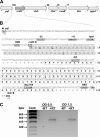
FIG. 1.
FIG. 1.
Structure and expression of the E. coli nrdR operon. (A) Organization of the genes. The nrdR operon comprises nrdR (ybaD), coding for NrdR, a transcriptional regulator of RNR genes; ribD, coding for the bifunctional diaminohydroxyphosphoribosylaminopyrimidine deaminase/5-amino-6-(5-phosphoribosylamino) uracil reductase; ribH, coding for the beta subunit of riboflavin synthase or 6,7-dimethyl-8-ribityllumazine synthase; nusB, coding for the transcription antitermination factor NusB; thiL, coding for thiamine-monophosphate kinase; and pgpA, coding for phosphatidylglycerophosphatase A. The arrows indicate the orientations of genes. (B) Nucleotide sequence of the nrdR regulatory region and structural gene. The deduced NrdR amino acid sequence is shown. The predicted zinc finger DNA binding motif and ATP cone motif are shown in shaded and open boxes, respectively. Predicted promoter −10 and −35 recognition elements, ribosomal binding site (RBS), and translational initiation and termination codons are shown in boldface. The arrows indicate the directions of transcription of genes. The restriction sites used in the construction of nrdR deletion mutants (see Materials and Methods) are shown boldface italics and underlined. (C) Semiquantitative RT-PCR analysis of nrdR at exponential (OD = 0.5) and early stationary (OD = 1.5) phases of growth. RT, reverse transcriptase.
FIG. 2.
FIG. 2.
Real-time RT-PCR analysis of nrdAB, nrdHIEF, and nrdDG in MG1655 (wild type), MG1655ΔnrdR1, and MG1655ΔnrdR2 RNAs as a function of the growth phase. Real-Time PCR measurements were performed using TaqMan primers and probes, and detection was performed in a model 7000 ABI Prism Sequence Detection System from Applied Biosystems. The induction factors in the two mutant strains are the ratios of the amount of specific RNA in the mutant strain to that in the wild type at the same OD normalized with the endogenous reference (gapA). (Top) nrdA probe. (Middle) nrdE probe. (Bottom) nrdD probe. Open bars, MG1655 wild type; black bars, MG1655ΔnrdR1; gray bars, MG1655ΔnrdR2.
FIG. 3.
FIG. 3.
Alignment of the E. coli (ECO) and S. enterica serovar Typhimurium (STY) nrdAB, nrdHIEF, and nrdDG regulatory regions showing the positions and sequences of the promoter elements and NrdR boxes. NrdR box motifs are aligned and are shown in boldface and enclosed in rectangular boxes. Identical bases in motifs are indicated by asterisks. The reported experimentally determined promoter −10 and −35 recognition elements are shown (8, 25, 47) in italics and underlined. DnaA box motifs are shown in boldface in white letters in gray boxes, and their relative orientations are indicated by arrows. A FUR box located upstream of the nrdHIEF genes is shown in boldface in a gray box. Two FNR box motifs in the upstream region of nrdD are shown in boldface and doubly underlined. Other symbols shown in boldface are tss, the transcriptional start site (where known), and the translational start codon.
FIG. 4.
FIG. 4.
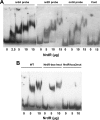
NrdR binding to the DNA regulatory region of the E. coli nrdAB, nrdHIEF, and nrdDG operons. DIG labeling of DNA probes and gel electrophoretic mobility shift assays were performed as described in Materials and Methods. (A) nrdA probe with 0, 2.5, 5, 10, and 15 μg protein; nrdD probe with 0, 5, 10, and 15 μg protein; nrdH probe with 0, 5, 10, and 15 μg protein; and control (Cont) rib probe with 0 and 15 μg protein. (B) NrdR binding to DNA of nrdA wild-type and mutant NrdR box 1 and box 2 probes. The NrdR box1 sequence was changed from 5′-TCACACTATCTTGCAG to 5′-TgAgACataCaTcCAG; the sequence of NrdR box 2 was changed from 5′-CCCCTATATATAGTGT to 5′-CgCgTAataAaAcTGT. The seven changes are shown in lowercase letters. Wild-type (WT) probe with 0, 5, and 10 μg protein; mutant (mut) NrdR box 1 probe with 0, 5, and 10 μg protein; mutant NrdR box 2 probe with 0, 5, and 10 μg protein.

Similar articles

Cited by

References

    1. Aravind, L., Y. I. Wolf, and E. V. Koonin. 2000. The ATP-cone: an evolutionarily mobile, ATP-binding regulatory domain. J. Mol. Microbiol. Biotechnol. 2:191-194. - PubMed
    1. Atkin, C. L., L. Thelander, P. Reichard, and G. Lang. 1973. Iron and free radical in ribonucleotide reductase. Exchange of iron and Mossbauer spectroscopy of the protein B2 subunit of the Escherichia coli enzyme. J. Biol. Chem. 248:7464-7472. - PubMed
    1. Baba, T., T. Ara, M. Hasegawa, Y. Takai, Y. Okumura, M. Baba, K. A. Datsenko, M. Tomita, B. L. Wanner, and H. Mori. 2006. Construction of Escherichia coli K-12 in-frame, single-gene knockout mutants: the Keio collection. Mol. Syst. Biol 2:2006.0008. - PMC - PubMed
    1. Blattner, F. R., G. Plunkett III, C. A. Bloch, N. T. Perna, V. Burland, M. Riley, J. Collado-Vides, J. D. Glasner, C. K. Rode, G. F. Mayhew, J. Gregor, N. W. Davis, H. A. Kirkpatrick, M. A. Goeden, D. J. Rose, B. Mau, and Y. Shao. 1997. The complete genome sequence of Escherichia coli K-12. Science 277:1453-1474. - PubMed
    1. Borovok, I., B. Gorovitz, R. Schreiber, Y. Aharonowitz, and G. Cohen. 2006. Coenzyme B12 controls transcription of the Streptomyces class Ia ribonucleotide reductase nrdABS operon via a riboswitch mechanism. J. Bacteriol. 188:2512-2520. - PMC - PubMed

Publication types

MeSH terms

Substances