. 2007 Jul;189(14):5293-301.
doi: 10.1128/JB.00234-07. Epub 2007 May 11.

A functional homing endonuclease in the Bacillus anthracis nrdE group I intron

Affiliations

A functional homing endonuclease in the Bacillus anthracis nrdE group I intron

David Nord et al. J Bacteriol. 2007 Jul.

Abstract

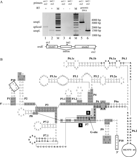
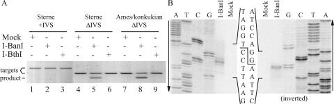
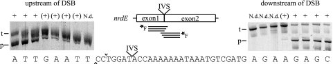
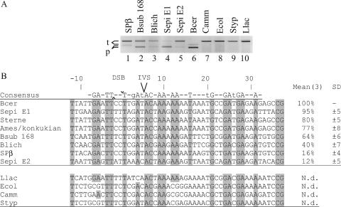
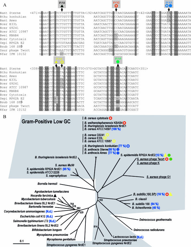
The essential Bacillus anthracis nrdE gene carries a self-splicing group I intron with a putative homing endonuclease belonging to the GIY-YIG family. Here, we show that the nrdE pre-mRNA is spliced and that the homing endonuclease cleaves an intronless nrdE gene 5 nucleotides (nt) upstream of the intron insertion site, producing 2-nt 3' extensions. We also show that the sequence required for efficient cleavage spans at least 4 bp upstream and 31 bp downstream of the cleaved coding strand. The position of the recognition sequence in relation to the cleavage position is as expected for a GIY-YIG homing endonuclease. Interestingly, nrdE genes from several other Bacillaceae were also susceptible to cleavage, with those of Bacillus cereus, Staphylococcus epidermidis (nrdE1), B. anthracis, and Bacillus thuringiensis serovar konkukian being better substrates than those of Bacillus subtilis, Bacillus lichenformis, and S. epidermidis (nrdE2). On the other hand, nrdE genes from Lactococcus lactis, Escherichia coli, Salmonella enterica serovar Typhimurium, and Corynebacterium ammoniagenes were not cleaved. Intervening sequences (IVSs) residing in protein-coding genes are often found in enzymes involved in DNA metabolism, and the ribonucleotide reductase nrdE gene is a frequent target for self-splicing IVSs. A comparison of nrdE genes from seven gram-positive low-G+C bacteria, two bacteriophages, and Nocardia farcinica showed five different insertion sites for self-splicing IVSs within the coding region of the nrdE gene.


Figures

FIG. 1.
FIG. 1.
Efficient splicing of the group I self-splicing intron in B. anthracis nrdE. (A) Agarose gel showing RT-PCR products of unspliced and spliced nrdE mRNA in total-RNA extraction from B. anthracis Sterne 7700 pXO1/pXO2. Exon1- and exon2-specific primers (ex1 and ex2, respectively) in RT-PCR gave a product of 2.1 kbp, corresponding to spliced nrdE mRNA (lane 1). The intron-specific primer int1, in combination with ex2, gave an RT-PCR product of 1.5 kbp, corresponding to unspliced nrdE mRNA (lane 2). Primer pairs were used in control RT-PCR without RT and showed no DNA contamination in the RNA extraction (lanes 3 and 4). Primer pairs were also used in PCRs with genomic DNA as primer controls and showed products of 3.2 kbp and 1.5 kbp, respectively (lanes 5 and 6). A molecular size marker (M) was run for reference, where bands 4 to 6, 8, and 10 from the bottom correspond to 1,000 bp, 1,500 bp, 2,000 bp, 3,000 bp, and 4,000 bp, respectively. (B) Predicted secondary structure of the nrdE intron. The lowercase letters indicate the coding sequence of the nrdE gene. The uppercase letters indicate the intronic sequence. Boldface uppercase letters indicate the ORF in the intron, with the numbers in the loop in P6.2 representing the sequences of B. thuringiensis serovar konkukian and B. anthracis Sterne. Conserved sequence elements (R and S), conserved base-paired regions (P1 to P9), and additional pairings (P3.1, P3.2, P5.1, P5.1a, P5.2, p5.2a, P6a, P6.1a to P6.1c, P6.2, P7.1, and P7.2) are shown. Alignment between the 5′ and 3′ splice sites can be promoted by the boxed nucleotides, UUGGU, in the P1 loop, and ACCAA, near P9, making pairing P10. The shaded boxes represent nucleotides identical with the group I intron in the recA gene in B. anthracis (17).
FIG. 2.
FIG. 2.
The B. anthracis nrdE intron encodes a GIY-YIG HEase. (A) I-BanI, identified as a GIY-YIG endonuclease, contains an N-terminal GIY-YIG domain (red box) with the GIY-YIG endonuclease motif, a C-terminal GIY-YIG domain (blue box) with a minor-groove DNA binding α-helix motif, and a helix-turn-helix motif (yellow box). A large deletion in I-BthI spans most of both the N-terminal and C-terminal domains. (B) [35S]Met-radiolabeled in vitro translation of I-BthI and I-BanI showed products of 5 kDa and 30 kDa, respectively.
FIG. 3.
FIG. 3.
Mapping of the cleavage site of the I-BanI HEase. (A) Agarose gel showing products from a cleavage assay with fluorescein-labeled targets, wild-type Sterne (2,276 bp; lanes 1 to 3), Sterne IVS-less (1,174 bp; lanes 4 to 6), and Ames/serovar konkukian (1,174 bp; lanes 7 to9) incubated with mock translation, I-BanI, and I-BthI. No cleavage products were found for targets incubated with mock translation or I-BthI (lanes 1, 4, and 7 and lanes 3, 6, and 9, respectively). Cleavage products corresponding to the predicted 722 bp were found for IVS-less targets incubated with I-BanI (lanes 5 and 8). No cleavage product was found for wild-type Sterne incubated with I-BanI (lane 2). Note that only one cleavage product was visualized, as only one strand was fluorescein labeled. (B) Polyacrylamide gel showing sequencing reactions run alongside 32P single-strand-labeled cleavage reactions to identify the I-BanI cleavage site and overhang. The second gel is inverted to more clearly show the cleavage sites on both strands.
FIG. 4.
FIG. 4.
Mapping of minimum sequence required for I-BanI cleavage. Polyacrylamide gels show products from cleavage assays with fluorescein-labeled targets with short flanking sequences upstream and downstream of the cut on the coding strand, −10 bp and + 35 bp, respectively. The target (t) and product (p) are indicated for each gel. Note that only one cleavage product was visualized, as only one strand was fluorescein labeled. The left and right gels show cleavage of the targets with successively shorter sequence upstream and downstream. The end positions are indicated below the lanes. Cleavage is indicated with + for efficient cleavage, (+) for low or nearly no cleavage, and N.d. for no cleavage detected. DSB is indicated with ^ and for the cuts on the template and coding strands, respectively.
FIG. 5.
FIG. 5.
I-BanI can cleave several closely related Bacillaceae nrdE genes. (A) Agarose gel showing products (p) from a cleavage assay with fluorescein-labeled targets (t) from the B. subtilis 168 prophage SPβ (2,106 bp), B. subtilis 168 (2,103 bp), B. licheniformis (2,103 bp), S. epidermidis nrdE1 (2,103 bp) and nrdE2 (2,103 bp), B. cereus (1,174 bp), C. ammoniagenes (2,106 bp), E. coli (2,106 bp), S. enterica serovar Typhimurium (2,106 bp), and L. lactis (2,106 bp) incubated with I-BanI. Cleavage products corresponding to the predicted sizes were found for targets B. subtilis 168 prophage SPβ (1,073 bp), B. subtilis 168 (784 bp), B. licheniformis (784 bp), S. epidermidis nrdE1 (787 bp) and nrdE2 (766 bp), and B. cereus (722 bp) (lanes 1 to 6). No cleavage products were detected for the targets C. ammoniagenes, E. coli, S. enterica serovar Typhimurium, and L. lactis (lanes 7 to 10). Note that only one cleavage product was visualized, as only one strand was fluorescein labeled. (B) Target site alignments showing a region from positions −12 to +37 flanking the cuts on the coding strands of all target sequences tested. For each target, the measured relative activity is indicated, with the most efficiently cleaved target set as 100%. N.d. indicates no cleavage detected. Means and standard deviations (SD) were calculated from measurements from three experiments. DSB is indicated with ^ and for the cuts on the template and coding strands, respectively. Uppercase letters in the consensus sequence and gray boxes indicate fully conserved nucleotides within all cleaved targets. Lowercase letters in the consensus sequence indicate conserved nucleotides in target sequences cleaved 50% or more.
FIG. 6.
FIG. 6.
The nrdE gene in Bacillaceae is a refuge for self-splicing IVSs. (A) Alignments of sequences flanking the insertion sites of IVSs in the nrdE gene (IVS1 to -5 in different colors) found within the family Bacillaceae, S. epidermidis, Staphylococcus phage Twort, and N. farcinica. The insertion sites of the IVSs and fully conserved nucleotides are indicated with light gray and dark gray, respectively. The numbers indicate the distances in nucleotides between the shown aligned sequence locations in the B. anthracis Sterne nrdE. Symbols denote group I intron with HEG (filled circles), group I intron without HEG (open circles), and intein with HEG (filled triangles). Inteins IVS1 and IVS4 have also been called RIR1-j and RIR1-b, respectively (http://bioinfo.weizmann.ac.il/∼pietro/inteins/), and introns IVS2, IVS3, and IVS4 have also been called RIR1-I, RIR1-II, and RIR1-III, respectively (20). (B) Phylogenetic tree of bacterial NrdEs with a selected set of Bacillaceae. The low-G+C gram-positive bacteria are shown with a shaded background. Vertical lines indicate subgroups within the B. cereus group, underlining denotes phage sequences, and dashed underlining denotes prophage. The occurrence of IVS1 to IVS5 is indicated with symbols and colors as shown in panel A, and relative cleavage efficiencies by I-BanI according to Fig. 4 are indicated. Only bootstrap values below 950 are shown. The distance bar in the lower left corner indicates 0.1 changes per site.

Similar articles

Cited by

References

    1. Belfort, M., and R. J. Roberts. 1997. Homing endonucleases: keeping the house in order. Nucleic Acids Res. 25:3379-3388. - PMC - PubMed
    1. Cech, T. R. 1990. Self-splicing of group I introns. Annu. Rev. Biochem. 59:543-568. - PubMed
    1. Chen, I., P. J. Christie, and D. Dubnau. 2005. The ins and outs of DNA transfer in bacteria. Science 310:1456-1460. - PMC - PubMed
    1. Chen, I., and D. Dubnau. 2004. DNA uptake during bacterial transformation. Nat. Rev. Microbiol. 2:241-249. - PubMed
    1. Claverys, J. P., M. Prudhomme, and B. Martin. 2006. Induction of competence regulons as a general response to stress in gram-positive bacteria. Annu. Rev. Microbiol. 60:451-475. - PubMed

Publication types

MeSH terms