. 2007 Jul;144(3):1316-27.
doi: 10.1104/pp.107.098152. Epub 2007 May 11.

The Flaveria bidentis beta-carbonic anhydrase gene family encodes cytosolic and chloroplastic isoforms demonstrating distinct organ-specific expression patterns

Affiliations

The Flaveria bidentis beta-carbonic anhydrase gene family encodes cytosolic and chloroplastic isoforms demonstrating distinct organ-specific expression patterns

Sasha G Tetu et al. Plant Physiol. 2007 Jul.

Abstract

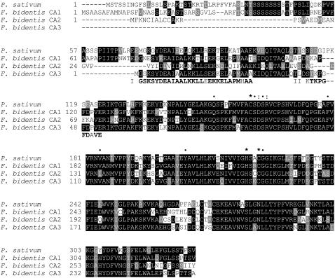
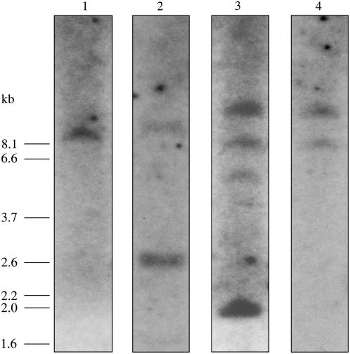
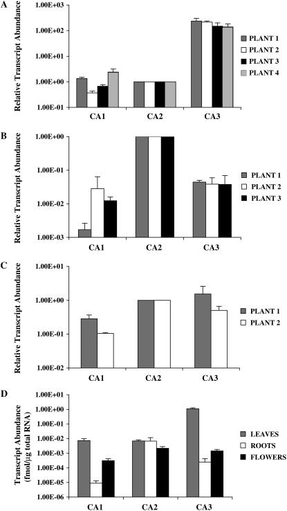
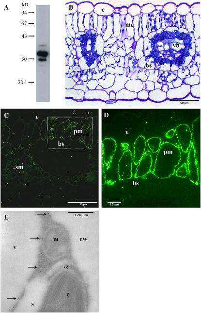
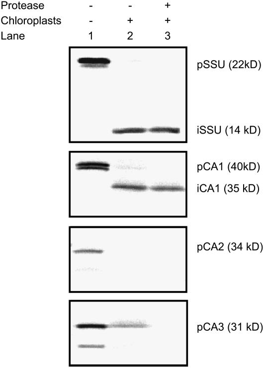
Carbonic anhydrase (CA) catalyzes the interconversion of CO(2) and bicarbonate, the forms of inorganic carbon used by the primary carboxylating enzymes of C(3) and C(4) plants, respectively. Multiple forms of CA are found in both photosynthetic subtypes; however, the number of isoforms and the location and function of each have not been elucidated for any single plant species. Genomic Southern analyses showed that the C(4) dicotyledon Flaveria bidentis 'Kuntze' contains a small gene family encoding beta-CA and cDNAs encoding three distinct beta-CAs, named CA1, CA2, and CA3, were isolated. Quantitative reverse transcription-polymerase chain reactions showed that each member of this beta-CA family has a specific expression pattern in F. bidentis leaves, roots, and flowers. CA3 transcripts were at least 50 times more abundant than CA2 or CA1 transcripts in leaves. CA2 transcripts were detected in all organs examined and were the most abundant CA transcripts in roots. CA1 mRNA levels were similar to those of CA2 in leaves, but were considerably lower in roots and flowers. In vitro import assays showed CA1 was imported into isolated pea (Pisum sativum) chloroplasts, whereas CA2 and CA3 were not. These results support the following roles for F. bidentis CAs: CA3 is responsible for catalyzing the first step in the C(4) pathway in the mesophyll cell cytosol; CA2 provides bicarbonate for anapleurotic reactions involving nonphotosynthetic forms of phosphoenolpyruvate carboxylase in the cytosol of cells in both photosynthetic and nongreen tissues; and CA1 carries out nonphotosynthetic functions demonstrated by C(3) chloroplastic beta-CAs, including lipid biosynthesis and antioxidant activity.


Figures

Figure 1.
Figure 1.
Alignment of the deduced amino acid sequences of pea CA and F. bidentis CA1, CA2, and CA3, and CA3 N-terminal amino acid sequences. Identical residues are boxed in black, whereas conservative changes are shown in gray. Dashes represent gaps inserted to maximize the alignment. Fragments obtained from N-terminal amino acid sequencing, I and II, are shown aligned in bold, except at mismatched residues. *, Residues implicated in Zn2+ binding; •, highly conserved active site residues of higher plant β-CAs; :, D/R pair found thus far in all β-CAs (Provart et al., 1993; Bracey et al., 1994; Kimber and Pai, 2000). GenBank accession number: pea, M63627.
Figure 2.
Figure 2.
Southern-blot analysis of F. bidentis genomic DNA. Genomic DNA was digested with XbaI, fragments were separated by electrophoresis, transferred to membrane, and then hybridized with radiolabeled CA1 ORF (lane 1), CA2 ORF (lane 2), CA3 ORF (lane 3), or the 118-bp CA3 exon probe (lane 4). Molecular size markers are shown at the left (in kb).
Figure 3.
Figure 3.
CA transcript abundance in F. bidentis organs. Relative transcript levels of the CA1, CA2, and CA3 genes in leaves (A), roots (B), and flowers (C) of individual plants. Values for CA1 and CA3 transcripts were normalized to those of the CA2 gene. Absolute CA transcript levels (D) in F. bidentis organs based on the total RNA used in the qRT-PCRs. A to C, sd of replicates done with the same cDNA preparation from an individual plant. D, se of the combined data from the individual plants shown in A to C.
Figure 4.
Figure 4.
Immunolabeling of F. bidentis CA isoforms. A, F. bidentis leaf proteins were separated by 12% SDS-PAGE, transferred to nitrocellulose, and labeled with an affinity-purified anti-F. bidentis CA3 antiserum. Immunoreactive proteins of approximately 35, 32, 30, and 28 kD were detected. Molecular mass markers shown in kD. B, A transverse section through a leaf of F. bidentis stained with methylene blue illustrates the characteristic Kranz anatomy of this C4 dicot. Vascular bundles (vb) are surrounded by bundle-sheath (bs) cells, which are surrounded by mesophyll cells (mc). The densely stained structures at the periphery of the mc and in a centripetal location in the bs are chloroplasts. e, Epidermal cell. C, An F. bidentis leaf in transverse section labeled with the affinity-purified anti-F. bidentis CA3 antiserum. Immunofluorescence was detected in the palisade (pm) and spongy (sm) mesophyll cells. Only nonspecific background fluorescence was seen in epidermal (e) and bundle-sheath (bs) cells. D, An immunolabeled adjacent section of the palisade mesophyll (pm) cells boxed in C at higher magnification. Bright fluorescence was seen at the periphery of the cells, corresponding to the location of the cytoplasm. Little or no fluorescence was detected in the epidermal (e) or bundle-sheath (bs) cells. E, Transverse section through part of a F. bidentis mesophyll cell labeled with the affinity-purified anti-F. bidentis CA3 antiserum followed by a secondary antibody coupled to colloidal gold particles. Gold particles (arrows) labeled the cytosol of the cells, and the chloroplast stroma (arrowhead). Mitochondria (m), starch grains (s), vacuole (v), and cell wall (cw) were not labeled.
Figure 5.
Figure 5.
Chloroplast import assays of in vitro transcribed and translated F. bidentis CA1, CA2, and CA3. Lane 1, Precursor (p) proteins generated for CA1, CA2, CA3, and the pea Rubisco SSU, which served as an import control. A single precursor was obtained for CA2, whereas two polypeptides were generated for the other constructs due to initiation of translation at internal Met residues as well as at the N termini. Lane 2, Chloroplast fractions following incubation of the precursor proteins under conditions favoring import. Imported (i) polypeptides were detected for CA1 and the SSU. Lane 3, As for lane 2, except protease was externally added to the chloroplasts prior to their collection.

Similar articles

Cited by

References

    1. Arnon DI (1949) Copper enzymes in isolated chloroplasts: polyphenoloxidase in Beta vulgaris. Plant Physiol 24 1–15 - PMC - PubMed
    1. Atkins C, Smith P, Mann A, Thumfort P (2001) Localization of carbonic anhydrase in legume nodules. Plant Cell Environ 24 317–326
    1. Atkins CA (1974) Occurrence and some properties of carbonic anhydrases from legume root nodules. Phytochemistry 13 93–98
    1. Bläsing OE, Ernst K, Streubel M, Westhoff P, Svensson P (2002) The non-photosynthetic phosphoenolpyruvate carboxylases of the C4 dicot Flaveria trinervia—implications for the evolution of C4 photosynthesis. Planta 215 448–456 - PubMed
    1. Bracey MH, Christiansen J, Tovar P, Cramer SP, Bartlett SG (1994) Spinach carbonic anhydrase: investigation of the zinc-binding ligands by site-directed mutagenesis, elemental analysis, and EXAFS. Biochemistry 33 13126–13131 - PubMed

Publication types

Associated data

LinkOut - more resources