

Restructuring of the dinucleotide-binding fold in an NADP(H) sensor protein
- PMID: 17496144
- PMCID: PMC1885584
- DOI: 10.1073/pnas.0700480104
Restructuring of the dinucleotide-binding fold in an NADP(H) sensor protein
Abstract
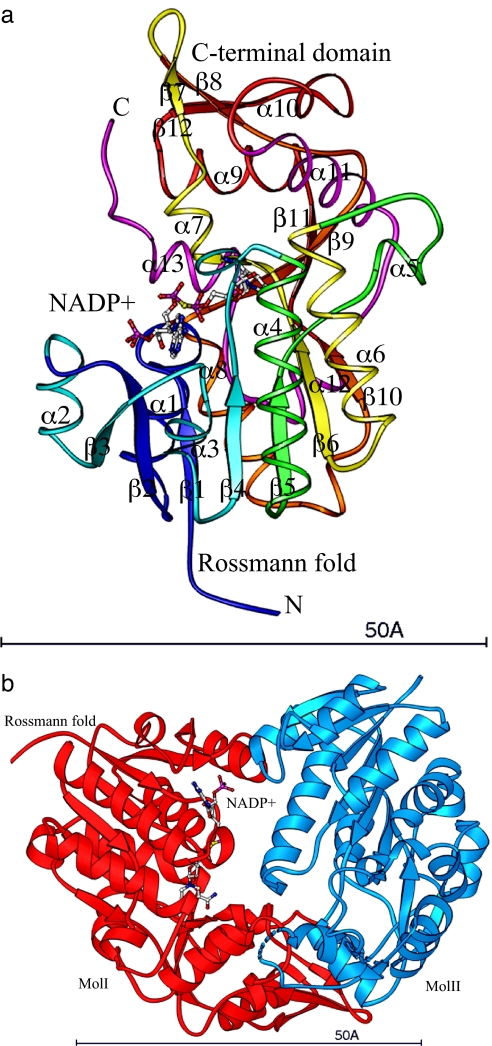
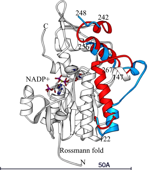
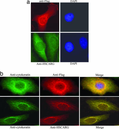
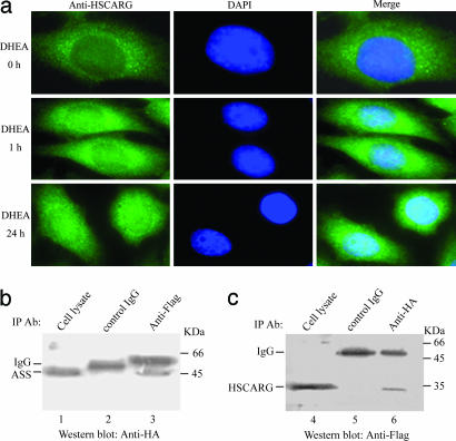
NAD(P) has long been known as an essential energy-carrying molecule in cells. Recent data, however, indicate that NAD(P) also plays critical signaling roles in regulating cellular functions. The crystal structure of a human protein, HSCARG, with functions previously unknown, has been determined to 2.4-A resolution. The structure reveals that HSCARG can form an asymmetrical dimer with one subunit occupied by one NADP molecule and the other empty. Restructuring of its NAD(P)-binding Rossmann fold upon NADP binding changes an extended loop to an alpha-helix to restore the integrity of the Rossmann fold. The previously unobserved restructuring suggests that HSCARG may assume a resting state when the level of NADP(H) is normal within the cell. When the NADP(H) level passes a threshold, an extensive restructuring of HSCARG would result in the activation of its regulatory functions. Immunofluorescent imaging shows that HSCARG redistributes from being associated with intermediate filaments in the resting state to being dispersed in the nucleus and the cytoplasm. The structural change of HSCARG upon NADP(H) binding could be a new regulatory mechanism that responds only to a significant change of NADP(H) levels. One of the functions regulated by HSCARG may be argininosuccinate synthetase that is involved in NO synthesis.
Conflict of interest statement
The authors declare no conflict of interest.
Figures
Similar articles
-
Structure and functions of cellular redox sensor HSCARG/NMRAL1, a linkage among redox status, innate immunity, DNA damage response, and cancer.Free Radic Biol Med. 2020 Nov 20;160:768-774. doi: 10.1016/j.freeradbiomed.2020.09.016. Epub 2020 Sep 17. Free Radic Biol Med. 2020. PMID: 32950687 Free PMC article. Review.
-
NADPH is an allosteric regulator of HSCARG.J Mol Biol. 2009 Apr 17;387(5):1277-85. doi: 10.1016/j.jmb.2009.02.049. Epub 2009 Feb 28. J Mol Biol. 2009. PMID: 19254724
-
An NADPH sensor protein (HSCARG) down-regulates nitric oxide synthesis by association with argininosuccinate synthetase and is essential for epithelial cell viability.J Biol Chem. 2008 Apr 18;283(16):11004-13. doi: 10.1074/jbc.M708697200. Epub 2008 Feb 8. J Biol Chem. 2008. PMID: 18263583
-
A structurally conserved water molecule in Rossmann dinucleotide-binding domains.Protein Sci. 2002 Sep;11(9):2125-37. doi: 10.1110/ps.0213502. Protein Sci. 2002. PMID: 12192068 Free PMC article.
-
NAD-binding domains of dehydrogenases.Curr Opin Struct Biol. 1995 Dec;5(6):775-83. doi: 10.1016/0959-440x(95)80010-7. Curr Opin Struct Biol. 1995. PMID: 8749365 Review.
Cited by
-
Cold Conditioned: Discovery of Novel Alleles for Low-Temperature Tolerance in the Vavilov Barley Collection.Front Plant Sci. 2021 Dec 15;12:800284. doi: 10.3389/fpls.2021.800284. eCollection 2021. Front Plant Sci. 2021. PMID: 34975991 Free PMC article.
-
Bovine NMRAL2 Protein Blunts Nitric Oxide Production and Inflammatory Response in Mycobacterium bovis Infected Bovine Lung Epithelial Cells.Cells. 2024 Nov 24;13(23):1953. doi: 10.3390/cells13231953. Cells. 2024. PMID: 39682702 Free PMC article.
-
The SDR (short-chain dehydrogenase/reductase and related enzymes) nomenclature initiative.Chem Biol Interact. 2009 Mar 16;178(1-3):94-8. doi: 10.1016/j.cbi.2008.10.040. Epub 2008 Nov 5. Chem Biol Interact. 2009. PMID: 19027726 Free PMC article.
-
Structure and functions of cellular redox sensor HSCARG/NMRAL1, a linkage among redox status, innate immunity, DNA damage response, and cancer.Free Radic Biol Med. 2020 Nov 20;160:768-774. doi: 10.1016/j.freeradbiomed.2020.09.016. Epub 2020 Sep 17. Free Radic Biol Med. 2020. PMID: 32950687 Free PMC article. Review.
-
Stamp2 controls macrophage inflammation through nicotinamide adenine dinucleotide phosphate homeostasis and protects against atherosclerosis.Cell Metab. 2012 Jul 3;16(1):81-9. doi: 10.1016/j.cmet.2012.05.009. Epub 2012 Jun 14. Cell Metab. 2012. PMID: 22704678 Free PMC article.
References
-
- Berger F, Ramirez-Hernandez MH, Ziegler M. Trends Biochem Sci. 2004;29:111–118. - PubMed
-
- Ying W. Front Biosci. 2006;11:3129–3148. - PubMed
-
- Hekimi S, Guarente L. Science. 2003;299:1351–1354. - PubMed
-
- Lin SJ, Kaeberlein M, Andalis AA, Sturtz LA, Defossez PA, Culotta VC, Fink GR, Guarente L. Nature. 2002;418:344–348. - PubMed
Publication types
MeSH terms
Substances
Associated data
- Actions
LinkOut - more resources
Full Text Sources
Molecular Biology Databases
Miscellaneous
