. 2007 Nov 8;26(51):7240-50.
doi: 10.1038/sj.onc.1210533. Epub 2007 May 14.

Endoglin inhibits prostate cancer motility via activation of the ALK2-Smad1 pathway

Affiliations

Endoglin inhibits prostate cancer motility via activation of the ALK2-Smad1 pathway

C S Craft et al. Oncogene. .

Abstract

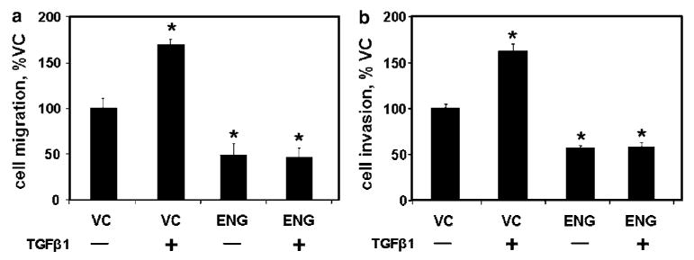
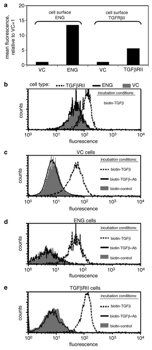
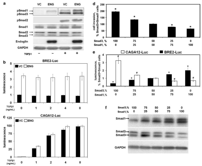
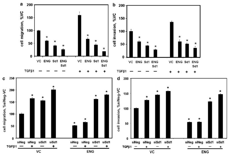
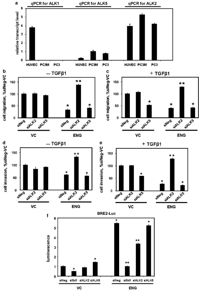
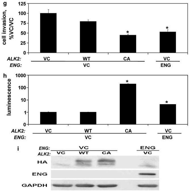
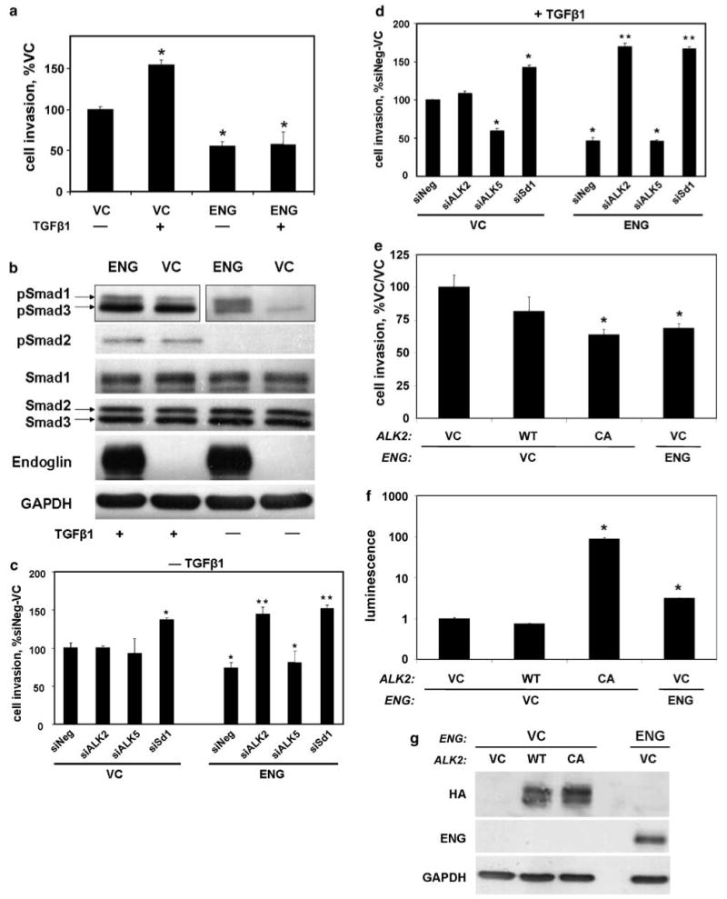
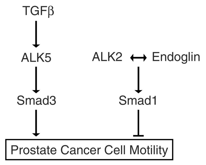
Endoglin is a transforming growth factor beta (TGFbeta) superfamily auxiliary receptor. We had previously shown that it suppressed prostate cancer (PCa) cell motility, and that its expression was lost during PCa progression. The mechanism by which endoglin inhibits PCa cell motility is unknown. Here we demonstrate that endoglin abrogates TGFbeta-mediated cell motility, but does not alter cell surface binding of TGFbeta. By measuring Smad-specific phosphorylation and Smad-responsive promoter activity, endoglin was shown to constitutively activate Smad1, with little-to-no effect upon Smad3. Knockdown of Smad1 increased motility and abrogated endoglin's effects. As type I activin receptor-like kinases (ALKs) are necessary for Smad activation, we went on to show that knockdown of ALK2, but not TGFbetaRI (ALK5), abrogated endoglin-mediated decreases in cell motility and constitutively active ALK2 was sufficient to restore a low-motility phenotype in endoglin deficient cells. These findings provide the first evidence that endoglin decreases PCa cell motility through activation of the ALK2-Smad1 pathway.


Figures

Figure 1
Figure 1
Endoglin (ENG)-mediated inhibition of cell motility is not altered by transforming growth factor β1 (TGFβ1). PC3-M cells were transfected with either ENG or with empty vector (VC) and β-galactosidase (β-gal) then treated with TGFβ1 (or not) for 24 h, as indicated. Cell migration (a) and invasion (b) were measured as described in the Materials and methods. Data are the mean±s.e.m. (N = 4) percent of VC cells from a single experiment; similar results were obtained in replicate experiments. (* denotes two-sided t-test P-value <0.05).
Figure 2
Figure 2
Endoglin (ENG) does not alter cell surface TGFβ1 binding. PC3-M cells were transfected with ENG, type II transforming growth factor β receptor (TGFβRII) or with empty vector (VC), as indicated. (a) Cell surface expression of ENG or TGFβRII was measured by FACS. Values represent the mean fluorescent intensity, normalized to the isotype control, from a single experiment. In (b), TGFβRII (broken line), ENG (solid line) or VC cells (solid gray) were incubated with biotinylated TGFβ1 (biotin-TGFβ), and, after washing, cell-associated biotin was measured by FACS analysis. Next, VC (c), ENG (d) or TGFβRII (e) cells were incubated with biotin-TGFβ (broken line), biotinylated negative control protein (biotin-control; solid gray) or with biotin-TGFβ pre-incubated with TGFβ-blocking antibody (biotin-TGFβ+Ab; solid line), and cell-associated biotin was measured by FACS analysis. Histograms from a single experiment are depicted. Cell transfection and FACS analysis were performed as described in the Materials and methods. All experiments were repeated with similar results.
Figure 3
Figure 3
Smad1 is activated by endoglin (ENG) and inhibits cell invasion. (a) ENG enhances phosphorylation of Smad1. PC3-M cells were transfected with ENG or VC, as in Figure 1. After treatment of cells with TGFβ1 (or not) for 30 min, resultant cell lysates were then probed by western blot for phosphorylated-Smad1, -2 and -3 (pSmad1, -2 and -3); total Smad1, -2 and -3 (Smad1, -2 and -3) and GAPDH, as described in the Materials and methods. Note: the pSmad1 and -3 blot for cells not treated with TGFβ1 was exposed longer than the TGFβ1 treated group in an attempt to detect pSmad3. (b and c) ENG enhances Smad1 promoter activity. Cells were co-transfected with ENG or VC, β-gal and BRE2-luciferase (BRE2-Luc) (b) or CAGA12-luciferase (CAGA12-Luc) (c). Six hours after changing to serum-free media, cells were treated with the indicated concentration of TGFβ1 for 18 h, and luciferase activity measured. VC cells not treated with TGFβ1 were normalized to 1.0 for each reporter construct. (d) Smad3 promotes cell invasion and Smad1 decreases it. Cells were transfected with the indicated ratios of Smad3 and -1, along with β-gal, and cell invasion measured. Data are the mean±s.e.m. (N = 4) of a single experiment, with similar results seen in a separate experiment (also N = 4). Compared to cells transfected with equal amounts of Smad3 and -1, * denotes values that differ by P<0.05 (two-sided t-test). The effects of different ratios of transfected Smad3 and -1, upon promoter activity (e; luciferase assay) and protein expression (f; western blot) are depicted. For promoter activity, cells were co-transfected with BRE2-Luc or CAGA12-Luc, as indicated. * and # denote values for BRE2-Luc and CAGA12-Luc, respectively, which differ from cells transfected with equal amounts of Smad3 and -1 with a P-value of <0.05. For all promoter assays, luciferase activity was normalized to β-gal; depicted values represent the mean±s.e.m. of two experiments performed on different days (N = 2 for each experiment).
Figure 4
Figure 4
Smad1 is necessary for endoglin (ENG)-mediated inhibition of cell motility. (a and b) ENG and Smad1 inhibit cell motility. PC3-M cells were transfected with VC, ENG, Smad1 (Sd1) or ENG and Sd1, then treated with TGFβ1 for 24 h, as indicated, and cell migration (a) and invasion (b) measured, as in Figure 1, and expressed as the percent of the corresponding VC cells. (c and d) Smad1 is necessary for ENG function. Cells were transfected with ENG or VC followed by either siRNA to Smad1 (siSd1) or nontargeting siRNA (siNeg), treated with TGFβ1 and cell migration (c) and invasion (d) measured, and expressed as the percent of non-TGFβ1-treated control cells (siNeg/VC). All values are the mean±s.e.m. (N = 4) of a single experiment, and repeated (also N = 4), with similar results. *P-value of <0.05, compared to control.
Figure 5
Figure 5
Activin receptor-like kinase 2 (ALK2) is necessary for endoglin (ENG) function. (a) Human prostate cancer cells express ALK2, but not ALK1. ALK1, ALK2 and ALK5 transcript levels were measured by quantitative reverse transcription (qRT)-PCR in PC3, PC3-M and human umbilical vein endothelial cells (HUVEC) cells (positive control), as described in the Materials and methods. Mean±s.d. relative transcript levels (normalized to glyceraldehyde-3-phosphate dehydrogenase (GAPDH)) are depicted. (be) ALK2 is necessary for ENG-mediated decreases in cell motility. PC3-M cells were transfected with ENG or empty-control vector (VC), along with β-gal, then with siALK2, siALK5 or siNeg, treated with TGFβ1 (c and e), or not (b and d) and cell migration (b and c) and invasion (d and e) measured. Data are the mean±s.e.m., expressed as percent siNeg/VC. (f) ALK2 supports ENG-mediated activation of Smad1. Cells were transfected with ENG or VC, along with β-gal and BRE2-Luc, and then with siSd1, siALK2, siALK5 or siNeg. Luciferase activity was measured (normalized to β-gal) and expressed as the mean±s.e.m. relative to VC-siNeg. (gi) Constitutively active ALK2 induces the ENG phenotype. Cells were transfected with HA-ALK2, HA-caALK2, ENG or VC, and cell invasion (g), luciferase activity (h) and HA-, ENG and GAPDH protein expression, by western blot (i), were measured. In (h), cells were co-transfected with β-gal and BRE2-Luc. Values are from a single experiment (replicates of N = 2 for qRT-PCR and luciferase; N = 4 for motility assays), and all experiments were repeated with similar findings. *P-value of <0.05, compared to siNeg/VC or to VC/VC (for G and H); **P-value of <0.05, compared to siNeg/ENG.
Figure 5
Figure 5
Activin receptor-like kinase 2 (ALK2) is necessary for endoglin (ENG) function. (a) Human prostate cancer cells express ALK2, but not ALK1. ALK1, ALK2 and ALK5 transcript levels were measured by quantitative reverse transcription (qRT)-PCR in PC3, PC3-M and human umbilical vein endothelial cells (HUVEC) cells (positive control), as described in the Materials and methods. Mean±s.d. relative transcript levels (normalized to glyceraldehyde-3-phosphate dehydrogenase (GAPDH)) are depicted. (be) ALK2 is necessary for ENG-mediated decreases in cell motility. PC3-M cells were transfected with ENG or empty-control vector (VC), along with β-gal, then with siALK2, siALK5 or siNeg, treated with TGFβ1 (c and e), or not (b and d) and cell migration (b and c) and invasion (d and e) measured. Data are the mean±s.e.m., expressed as percent siNeg/VC. (f) ALK2 supports ENG-mediated activation of Smad1. Cells were transfected with ENG or VC, along with β-gal and BRE2-Luc, and then with siSd1, siALK2, siALK5 or siNeg. Luciferase activity was measured (normalized to β-gal) and expressed as the mean±s.e.m. relative to VC-siNeg. (gi) Constitutively active ALK2 induces the ENG phenotype. Cells were transfected with HA-ALK2, HA-caALK2, ENG or VC, and cell invasion (g), luciferase activity (h) and HA-, ENG and GAPDH protein expression, by western blot (i), were measured. In (h), cells were co-transfected with β-gal and BRE2-Luc. Values are from a single experiment (replicates of N = 2 for qRT-PCR and luciferase; N = 4 for motility assays), and all experiments were repeated with similar findings. *P-value of <0.05, compared to siNeg/VC or to VC/VC (for G and H); **P-value of <0.05, compared to siNeg/ENG.
Figure 6
Figure 6
The activin receptor-like kinase 2 (ALK2)-Smad1 pathway is important for endoglin (ENG) (a) ENG inhibits cell invasion. PC3 cells were transfected with either ENG or with empty vector (VC), along with β-gal, and treated with transforming growth factor β1 (TGFβ1) (or not) for 24 h. Depicted are the mean±s.e.m. number of invading cells as a percent of no TGFβ1 vector control. (b) ENG increases Smad1 phosphorylation. Cells were transfected with either ENG or VC and treated with TGFβ (or not) for 30 min, and the resultant cell lysates were probed for the indicated phospho- and total protein species by western blot. Note the blot for pSmad1 and -3, for cells not treated with TGFβ1, was exposed longer than the TGFβ1 treated group in an attempt to detect pSmad3. (c and d) ENG-mediated inhibition of cell invasion is dependent upon ALK2 and Smad1. Cells were transfected as indicated, as in Figure 5, and treated with TGFβ (d) for 24 h, or not (c). Depicted are the mean±s.e.m. number of invading cells as a percent of siNeg/VC for a single experiment. (eg) Constitutively active ALK2 induces the ENG phenotype. Cells were transfected with HA-ALK2, HA-caALK2, ENG or VC, and cell invasion (e), luciferase activity; normalized to β-gal (f), and HA-, ENG and glyceraldehyde-3-phosphate dehydrogenase (GAPDH) protein expression, by western blot (g), were measured. In (f), cells were co-transfected with β-gal and BRE2-Luc. All experiments (N = 4 replicates for invasion) were repeated with similar results. *P-value <0.05, compared to VC (a), siNeg/VC (c and d) or VC/VC (e and f); **P-value of <0.05, compared to siNeg/ENG.
Figure 7
Figure 7
Model of endoglin (ENG)-mediated regulation of prostate cell motility.

Similar articles

Cited by

References

    1. Attisano L, Carcamo J, Ventura F, Weis FM, Massague J, Wrana JL. Identification of human activin and TGF beta type I receptors that form heteromeric kinase complexes with type II receptors. Cell. 1993;75:671–680. - PubMed
    1. Barbara NP, Wrana JL, Letarte M. Endoglin is an accessory protein that interacts with the signaling receptor complex of multiple members of the transforming growth factor-beta superfamily. J Biol Chem. 1999;274:584–594. - PubMed
    1. Bergan R, Hakim F, Schwartz GN, Kyle E, Cepada R, Szabo JM, et al. Electroporation of synthetic oligodeoxy-nucleotides: a novel technique for ex vivo bone marrow purging. Blood. 1996a;88:731–741. - PubMed
    1. Bergan R, Kyle E, Nguyen P, Trepel J, Ingui C, Neckers L. Genistein-stimulated adherence of prostate cancer cells is associated with the binding of focal adhesion kinase to beta-1-integrin. Clin Exp Metastasis. 1996b;14:389–398. - PubMed
    1. Bissell MJ, Radisky D. Putting tumours in context. Nat Rev Cancer. 2001;1:46–54. - PMC - PubMed

Publication types